Total Hip Arthroplasty: Direct Anterior Approach Using a Specialized Table
Introduction
Surgical approach first described by Carl Heuter in 1881, later adapted for arthroplasty and published by Robert Judet in 1950
Recent increase in popularity after series of anterior approach total hip arthroplasties published by Joel Matta in 2005
Further refinement has allowed for accelerated recovery and elimination of hip precautions
Potential downsides include steep learning curve, cost of a specialized table, and questioned benefits compared with modern posterior approach
Surgical Technique
| Video 58.1 Direct Anterior Approach Using a specialized table. Roy Davidovitch, MD; Dylan Lowe, BSc; Ran Schwarzkopf, MD, MSc (15 min 29 s) |
Patient Positioning and Preparation
Specialized radiolucent table that allows for sustained external rotation and extension of the surgical hip during preparation of the proximal femur
Fluoroscopic imaging per surgeon preference
Perineal post with no traction applied to the lower limbs
Antiseptic (chlorhexidine) preparation of the surgical site from umbilicus to knee. Drape the surgical hip as well as image intensifier
Surgical Anatomy and Approach

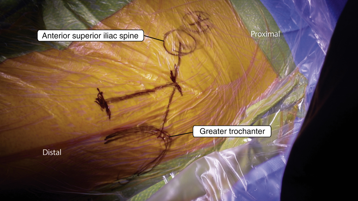
Figure 1Intraoperative photograph. The surgical incision is marked by a vertical line starting 2 cm lateral and 1 cm distal to the line drawn connecting the anterior superior iliac spine and the greater trochanter and drawn toward the lateral border of the patella.

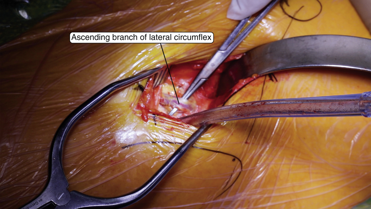
Figure 2Intraoperative photograph. The ascending branch of the lateral circumflex artery is identified.
The authors prefer a vertical incision; however, a “bikini” incision may be used
Mark the anterior superior iliac spine (ASIS) and greater trochanter and draw a line connecting the two
Incision begins 2 cm lateral and 1 cm distal to the ASIS and extends distally toward the lateral border of the patella. The typical incision is 8 to 10 cm in length (Figure 1)
Incision carried down to the level of the tensor fascia which is then incised
Fascia bluntly separated from the muscle and interval between tensor and rectus developed
Place a blunt cobra retractor over the superior femoral neck. The intermuscular interval between the tensor and sartorius can now be developed
Identify the ascending branch of the lateral femoral circumflex and cauterize (Figure 2)
Elevate the soft tissues off the anterior capsule and place a second blunt cobra retractor around the inferior neck
The capsule is incised in line with the center of the femoral neck followed by a capsulectomy of both the superior and inferior edges at the acetabular rim
Lateral capsule excised along the intertrochanteric line
The superior cobra retractor is placed intra-articular along the superior femoral neck
Stay updated, free articles. Join our Telegram channel

Full access? Get Clinical Tree


