CHAPTER SEVEN Exercise to Increase Range of Movement and Flexibility
This chapter discusses factors affecting normal and limited range of movement. Methods of assessing range of movement are considered. Principles of exercise design, exercise prescription for increasing range of movement and the training adaptations seen in response to a successful exercise programme are addressed.
DEFINITION
Range of movement (ROM) refers to the range through which the bones of a joint can be moved.
Active ROM refers to the range of joint movement produced by a voluntary muscle contraction. Passive ROM is the range through which the joint can be moved by the application of an external force, such as that produced by a physiotherapist. For some joints active and passive ROM may be different.
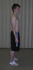
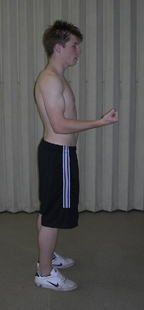
Range can also be described in terms of the excursion of the muscle producing the movement; the position when the muscle is in its shortest position is termed ‘inner range’ (Figure 7.1), and the position when the muscle is at its longest is termed ‘outer range’ (Figure 7.3), with ‘mid-range’ falling in between the two (Figure 7.2).
Flexibility can be defined as the ROM of a joint or joints, or alternatively the freedom to move.
It is important that joints have a balance between stability, which limits abnormal direction and range of movement, and flexibility, for ease of movement. The type and position of a joint will determine the amount of movement available at the joint, along with some of the other specific factors listed below.
FACTORS LIMITING NORMAL RANGE OF MOVEMENT
Ligaments around a joint
The length and tension of the ligaments limit joint movement as most ligaments are inelastic because they are made up of white fibrous collagen. When a ligament or tendon is at its full length no further movement in that direction is possible. In this way ligaments both restrict movement and direct the movement of the articulating surfaces over one another. An example of normal ROM, limited by ligaments, is the limitation of knee extension by the anterior cruciate ligament which prevents anterior sliding of the tibia on the femur.
FACTORS CAUSING ABNORMAL RANGE OF MOVEMENT
Immobility
Articular structures
After prolonged immobilization, 32 weeks, the articular structures become the main limiting factor to movement (Trudel 2000). The mechanism of intra-articular limitation is not clear, although the proliferation of intra-articular connective tissue, increase in collagen cross-linking and adaptive shortening of the capsule have all been suggested (Trudel 2000).
Muscle
The lack of longitudinal force through a muscle during immobilization leads to tissue remodelling to accommodate the new shortened resting length. Muscles immobilized in a shortened position over a period of time demonstrate a reduction in sarcomeres (Goldspink et al 1974), and an increase in the proportion of connective tissue which results in a decrease in joint ROM and compliance of the muscle (Williams 1988). These changes in sarcomeres can be seen as early as 24 hours after immobilization (McLachlan 1983). There is evidence from animal studies demonstrating that after a period of 2 weeks of immobilization the main factor limiting movement is muscle shortening.
Collagen
After a period of immobilization there is a change to the organization of the collagen within the muscle, leading to an increase in the number of perpendicularly orientated collagen fibres, which connect two adjacent muscle fibres (Jarvinen et al 2002). Chains of collagen molecules contain cross-links which weld them into a strong unit. It has been suggested that during immobilization there is a change in chemical structure and loss of water within the collagen which leads to the fibres coming into close contact with one another. This close contact is thought to lead to the formation of abnormal crossbridges, leading to an increase in tissue stiffness (Alter 2004).
Stay updated, free articles. Join our Telegram channel

Full access? Get Clinical Tree


