CHAPTER ONE Introduction to Exercise Physiology
This chapter provides a broad introduction to exercise physiology. Physiology of muscle, the cardiovascular system and energy supply are considered here. This chapter is not intended to be an in-depth study of this area but contains the underpinning knowledge with which physiotherapist who prescribes exercise in practice should be familiar. More details of any of the topics covered can be found in other exercise physiology texts and there are some suggestions of these at the end of the chapter.
MUSCLE
Skeletal muscle accounts for 40–50% of total body weight. It has three main functions:


Structure
Skeletal muscle is made up of fascicles or bundles of muscle fibres and is surrounded by and held together with connective tissue. This connective tissue forms three layers, the epimysium which surrounds whole muscles, the perymysium which surrounds fascicles or bundles of 10–100 muscle fibres and the endomysium which surrounds individual muscle fibres. The connective tissue layers hold the muscle together, connect muscle to other structures in the body and form tendons to connect muscle to bone. When a muscle contracts, tension is transmitted through the connective tissue which pulls on the muscle insertion and produces movement.
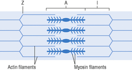
Muscle fibres are long cylindrical cells which lie parallel to one another. The plasma membrane of the muscle cell (sarcolemma) contains many myofibrils lying lengthways, within which are the contractile elements of the muscle. The myofibrils are composed of filaments, which are arranged in compartments called sarcomeres. These filaments are both thick and thin and overlap by differing amounts depending on whether the muscle is relaxed, contracting or stretched. This causes the striated (alternate light and dark bands) appearance of skeletal muscle at microscopic level. The component parts of a sarcomere are illustrated in Figure 1.1. Each sarcomere lies between Z lines. The A band is mostly made of myosin and does not change in length with contraction. The I band is mostly made of actin but there is also overlap of myosin here. This band alters in length with contraction.
Contraction
Muscles contract according to the sliding filament theory first described by Huxley in the 1960s. The thick filaments made up of the protein myosin have crossbridges which extend towards the thin filaments. These are formed mainly of the protein actin but also contain troponin and tropomyosin. Tropomyosin is attached to troponin. These are regulatory proteins to stop myosin and actin from making contact (Figure 1.2a). At the start of a muscle contraction, calcium attaches to troponin, changes its shape and moves the tropomyosin. The myosin heads (crossbridges) can then attach to the actin and pull on the thin filaments to generate force (Figure 1.2b). When a muscle relaxes tropomyosin again covers the myosin binding site.
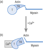
Figure 1.2 Protein crossbridges during muscle contraction and relaxation.
Adapted from Borell D, Nimmo M and Wood L (1996) Principles of Physiology. WB Saunders Ltd, p. 106
Skeletal muscle fibre types
Different types of muscle fibres have been described and there are many ways of classifying these according to functional characteristics. In Table 1.1 muscle fibre types are classified according to speed of contraction.
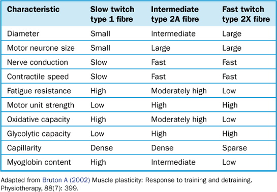
Most muscle groups within the body have an equal amount of type 1 and type 2 muscle fibres. Fifty per cent of the type 2 muscle fibres are 2a and 50% 2b. Some muscles have higher proportions of type 1 or type 2 fibres depending on the type of activity which they usually perform, e.g. postural muscles have a high proportion of type 1 fibres as they are used almost continuously throughout waking hours.


