Fig. 1.1
Schematic drawings for palmar view of the wrist joint complex showing the eight carpal bones and their articulations with the distal radius, distal ulna, the metacarpal bones of the hand and each other. H hamate, C capitates, TZ trapezoid, TP trapezium, TQ triquetrum, P pisiform, L lunate, S scaphoid. The arrows indicate the line of the joints. a carpometacarpal; b midcarpal; c radiocarpal. The light brown surfaces indicate the articular cartilages [8, 9]
The radiocarpal joint, which is arguably the most critical articulation in the wrist joint [2–4] consists of 75 % of the articulation between proximal carpal bones and the distal radius, whereas the remaining 25 % is the ulnacarpal or in particular, triangular fibrocartilage complex (TFCC) together with two main facets: elliptical scaphoid facet and spherical lunate facet [5]. There is a fibrocartilage ridge separating these two facets, known as the interfacet prominence. Concavity with declination 10–15o palmarly and 15–25o ulnarly is also observed at the distal articular surface of the radius [6]. TFCC, which functions as a buffer between the triquetrum and the distal tip of the ulna during ulnar deviation, consists of many components. Fibrocartilaginous disc, dorsal radioulnar and palmar ligaments, ulnocarpal ligaments, and the tendon sheath of the extensor carpi ulnaris form the TFCC [7]. Figure 1.2 illustrates the radiocarpal joint.
Fig. 1.2
The radiocarpal joint from distal [2]. Illustrated are the two fossae for the scaphoid and lunate bone
There are five main muscles associated with the motion of the wrist joint [8]. Radial deviation motion is performed by the extensor carpi radialis brevis, which is inserted onto the proximal end at the dorsal surface of the third metacarpal bone. Another tendon with the same function namely extensor carpi radialis longus is also inserted onto the proximal end at the dorsal surface of the second metacarpal bone. The last of the three muscles acting on the radial side, namely the flexor carpi radialis contributes to radial deviation and flexion of the wrist joint. The insertion is found at the palmar side of the base of the second metacarpal. The flexor and extensor carpi ulnaris muscles which are found at the ulnar side, flex and extend the wrist joint respectively. Both of them are acting together to perform ulnar deviation motion. There are three insertion points for the tendon of the flexor carpi ulnaris, where one of them is found at the pisiform. The remaining two are connected via ligaments onto the hamate and fifth metacarpal bone.
1.2 Bone Structure
Bones at the wrist joint have unique structure, and therefore names were given according to their shape or structure (Fig. 1.3). For instance, the name of the second largest carpal bone, the scaphoid is derived from the Greek “scaphe” which has the same meaning as “rowing boat” since it shaped like a boat [8].
As aforementioned, there are eight carpal bones where each of their names was given according to their shape. Table 1.1 provides information on the structure for each bone of the wrist joint.
Table 1.1
Description on the different structures of the eight carpal bones
Bone | Description |
|---|---|
Scophoid 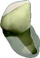 | The scaphoid can be divided into three main regions: the proximal pole, the waist and the distal pole. Several characteristics of the scaphoid have been identified, which include a single convex distal surface for articulation with the trapezium and trapezoid, a large concave distal capitate surface, a flat semilunar lunate surface medially and a large convex radial articular area extending dorsally [8] |
Lunate  | The next bone wedged between the scaphoid and triquetrum with its crescent shape is the lunate. This bone articulates laterally with the scaphoid via its flat semilunar facet. The medial part, on the other hand articulates with the triquetrum through its square surface. The radius, on its proximal region is articulated via convex facet. Distally, its convex surface articulates with the capitate |
Triquetrum 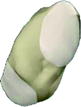 | The most medial bone of the proximal row carpal bone is the triquetrum. This small and irregularly shaped bone has distal concavo-convex surface which articulates with the hamate. At the anterior side of the bone, its oval convex palmar facet articulates with the pisiform whilst radially, the lunate is articulated with a square surface of the triquetrum |
Pisiform  | The sesamoid bone of the tendon of the flexor carpi ulnaris, which is the pisiform has a single flat oval articular facet. This facet allows articulation with the triquetrum on its dorsal surface [10] |
Hamate 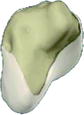 | The wedge shape bone, hamate has articular surfaces for the capitate and triquetrum on either sides. For articulation with the fifth and fourth metacarpals, there is a ridge at the distal articular surface, functions as a divider between medial and lateral facets. A hook on its palmar side, also known as hamulus, functions as an attachment point for certain ligaments. This criterion gives the hamate its distinct shape [8] |
Capitate 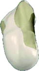 | The largest carpus namely the capitate which lies in the center of the wrist has a convex surface of its proximal part which is articulated with the lunate. Meanwhile, its lateral surface articulates with the trapezoid and scaphoid, and medial surface with the hamate. A concave distal strip on its lateral part is for articulation with the second metacarpal base whilst the third metacarpal base was articulated via a concavo-convex distal facet. The name of capitate is derived from the Latin word ‘capitãtus’ which means ‘containing head’ due to a rounded head-shaped on its surface |
Trapezoid  | A relatively smaller bone namely the trapezoid, on the other hand is articulated medially with the trapezium via a flat facet surface. On its proximal side, the scaphoid is articulated through a slightly concave surface whilst on its medial side, the capitate is articulated via a flat facet surface. Distally, the second metacarpal is articulated via a convex triangular surface [8] |
Trapezium 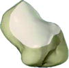 | The trapezium is located between the first metacarpal distally and the scaphoid proximally. For the articulation with the first metacarpal, it has a saddle shaped distal articular surface |
1.3 Cartilage Structure
Cartilage comprises of a dense network of collagen fibers embedded in a gel-like component of the ground substance namely chondroitin sulfate [11]. It lacks blood vessels, lymphatics and nerves [8]. There are three types of cartilages: hyaline cartilage, fibrocartilage and elastic cartilage. Articular cartilage which is composed from hyaline cartilage is mainly located at the ends of long bones. It promotes flexibility, support as well as smooth surfaces to assist movements of joints [11]. Four layers exist as shown in Fig. 1.4 with the topmost layer namely superficial zone which has the greatest importance. Since articular cartilage functions as a nearly frictionless bearing while uniformly transferring loads on underlying bone preventing high stress concentrations [12], this layer acts by supporting more than 90 % of the compressive loads. This corresponding surface also functions to resist shear forces generated by the joint movement. The fluid and collagen content in the cartilage differ, which subsequently lower in the intermediate, deep and calcified zones of articular cartilage. The collagen fibers determine its strength and its resilience, which is an ability to restore its original shape after deformation attributed to the presence of chondroitin sulfate.
Fig. 1.4
 Finite Element Modelling of the Healthy Wrist Joint
 The Wrist Joint Affected by Rheumatoid Arthritis
 Finite Element Analysis of the Wrist Arthroplasty in Rheumatoid Arthritis
 Biomechanical Properties and Behaviours of the Wrist Joint
 Finite Element Analysis of the Wrist Joint Affected by Rheumatoid Arthritis
 Malignant Brain Tumors


The articular cartilage which consists of four existing layers: superficial, intermediate, deep and calcified [19]
Related posts:
Finite Element Modelling of the Healthy Wrist Joint
The Wrist Joint Affected by Rheumatoid Arthritis
Finite Element Analysis of the Wrist Arthroplasty in Rheumatoid Arthritis
Biomechanical Properties and Behaviours of the Wrist Joint
Finite Element Analysis of the Wrist Joint Affected by Rheumatoid Arthritis
Malignant Brain Tumors
Stay updated, free articles. Join our Telegram channel
Full access? Get Clinical Tree
