Fig. 16.1
The ROBODOC System (Think Surgical, Inc., Fremont, CA)
Once the scan is complete, the CT data is used as input into ORTHODOC, which combines the individual slices to produce a series of 2D images for templating purposes. A 3D surface model of the operative bone is also created in ORTHODOC for intraoperative registration purposes. A sample ORTHODOC screen used for planning THA is shown in Fig. 16.2. ORTHODOC contains an open library of 510(k) cleared hip or knee replacement implants, depending on the application installed. The surgeon can select an implant model from this library and manipulate the 3D representation of the implant in relation to the bone to optimally place the implant. Once the surgeon is satisfied with the implant location, the data is written to a transfer media file for use with the ROBODOC during surgery.
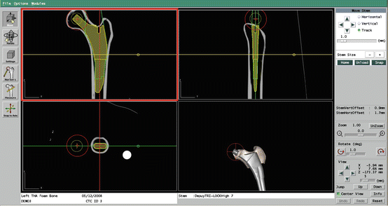
Fig. 16.2
An example of the 3D planning workstation, ORTHODOC, for a THA case
Technique
In the operating room, surgical exposure during a ROBODOC surgery is the same as is done during a normal TKA or THA. Once the joint is exposed, fixation is required to ensure that the operative bones are immobilized with respect to the ROBODOC base. Fixation is specific to the indication (i.e., THA fixation fixes the proximal femur while TKA fixation fixes the distal femur and proximal tibia). Once fixation has been established, bone motion recovery markers are placed on the bone. Recovery markers are used to recover the 3D location and orientation of the bone in the event that the bone moves after it has been registered. Bone motion monitors (BMM’s) are directly attached to the operative bones and can determine if a bone motion occurs. If a bone motion occurs during registration or cutting, the procedure is automatically paused by the system. This requires the bone be re-registered before the procedure may continue.
After the BMM’s have been attached, the next step is to register the operative bone within ROBODOC’s workspace. The current version of ROBODOC uses a widely accepted method of registration used in computer vision based on a point-to-surface technique. It requires the creation of a surface model of the bone from the preoperative CT scan using a semi-automated process in ORTHODOC and collecting, or digitizing, bone surface points on the patient intraoperatively.
During surgery, the surgeon uses a digitizer located on the ROBODOC system to collect points on the bone with respect to the ROBODOC arm coordinate system. The software directs the surgeon towards areas on the surface of the bone where points need to be collected. This method does not require the collection of specific anatomic points, but rather, the algorithm is designed to match the surface of the bone based on a variety of points in anatomic regions that are sufficiently spread out. If the points are not well distributed, a software check will require the points to be recollected until they are sufficiently spread apart. If the calculated registration meets the accuracy requirements, the surgeon is asked to verify the results of the registration by digitizing specific points. The graphic display then indicates where each collected point lies relative to the CT surface model. If they are acceptable to the surgeon, he/she can accept the registration and proceed with the surgery.
The next step of a ROBODOC procedure is the milling of the bone using a rotary cutting tool (Fig. 16.3). The surgeon must ensure that the soft tissue is properly retracted prior to letting ROBODOC begin cutting of the bone. During the entire bone preparation procedure (Fig. 16.4), the surgeon is in control of the process and is in direct view of the operative site while ROBODOC is cutting. The surgeon has the ability to pause, stop, or abort the use of ROBODOC at any point via a hand-held controller. During the entire ROBODOC procedure, the surgeon is guided through the workflow through on-screen prompts and displays.
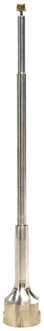
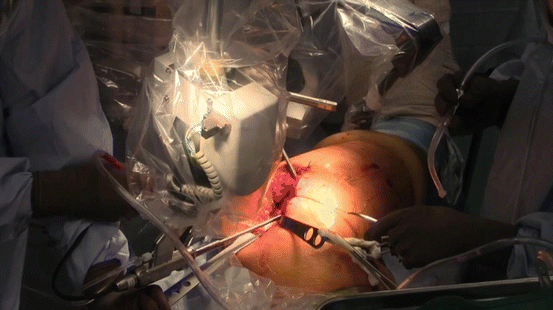
Fig. 16.3
An example of a ROBODOC THA cutting tool and sleeve used to prepare the bone surface
Fig. 16.4
ROBODOC preparing the femoral canal during THA
Once ROBODOC has finished preparing the bone, the software will prompt the surgeon to move ROBODOC away from the operating table. At this point, the surgeon removes the recovery markers and fixation system and proceeds with implant fitting and insertion in the same manner as a manual hip or knee arthroplasty. Finally, the exposed joint can be closed per standard procedures.
Results and Complications
The ROBODOC system has been used in thousands of clinical cases for both THA and TKA. A summary of the reports in which ROBODOC was used for THA and TKA are presented Tables 16.1 and 16.2, respectively. The reports and their primary findings are discussed in the sections below. It should be noted that all of these studies were performed with a variety of different implants created by different implant manufacturers.
Table 1
Clinical studies using ROBODOC for THA
Study | Procedure | No. cases | Blood loss (cc) | OR time (min) |
|---|---|---|---|---|
Bargar et al. (1998), US | THA | 65/62 | 1,189/644cca | 258/134a |
Bargar et al. (1998), Germany | THA | 900/– | – | 90/– |
Honl et al. (2003) | THA | 61/80 | – | 107/82a |
Nishihara et al. (2004) | THA | 75/– | – | – |
Nishihara et al. (2006) | THA | 78/78 | 527/694a | 122/102a |
Hananouchi et al. (2006) | THA | 31/27 | – | – |
Schulz et al. (2006) | THA | 143/– | – | – |
Nakamura et al. (2009) | THA | 75/75 | 591/– | 129/– |
Nakamura et al. (2010) | THA | 118/– | 120/108 | |
Yamamura et al. (2013) | RTHA | 19/– | 1,235 | 267 |
THA
As mentioned previously, the first human cases of ROBODOC were performed in 1992. Bargar et al. [2] describe the results of the first clinical trial using ROBODOC in the US along with the first 900 THA procedures performed in Germany. Under European law, all cases performed in Europe were performed after the system received CE mark. The US cohort was a controlled and randomized study with 65 ROBODOC cases and 62 manual control cases. There were no differences in functional outcomes in the two groups. Radiographic fit and component positioning was improved in the ROBODOC group but surgical time and blood loss were significantly greater in the ROBODOC group. The control group had three cases of femoral fracture while the ROBODOC group had none. In the German group, 870 of the cases were primary total hip arthroplasties with the remaining 30 bieng revision THA cases. The Harris hip scores rose from 43.7 preoperatively to 91.5 postoperatively and the learning curve for the system was demonstrated as the first case was 240 min, while the majority of cases were 90 min, on average. Complication rates were similar to conventional techniques, except the ROBODOC cases had no intraoperative femoral fractures.
Honl et al. [14] described their experiences using the ROBODOC system in 2003 in a randomized prospective clinical study. Of the 74 ROBODOC cases, 13 of them had to be converted to manual technique due to technical complications. Revision was required in two of the manual group and nine of the ROBODOC group. The ROBODOC cases took longer at 107.1 min on average, while the conventional cases only took 82.4 min on average. The ROBODOC group had significant improvements in limb-length equality and varus-valgus orientation of the stem. Additionally, the ROBODOC group had more heterotopic ossification at 6 months along with better Mayo clinical scores and Harris scores at 12 months. However, dislocation was higher in the ROBODOC group with 11 of the 61 cases reporting dislocation and eight of them requiring revision surgery compared to none in the conventional group. It should be noted that all of the revisions were for recurrent dislocations and pronounced limp. At revision, the authors found the abductor muscles were detached from the trochanter and implied that in those cases, the robot damaged the abductor muscles, causing rupture. Since ROBODOC is designed such that the tool shall not deviate from the prescribed cut path, it appears as though there were issues with properly clearing the workspace prior to allowing ROBODOC to cut. These results were unrepresentative of what was found in nearly all other reports. When the revision cases were excluded, the authors found the Harris hip scores, prosthetic alignment, and limb length differentials were better for the ROBODOC group at both 6 and 12 months.
In 2004, Nishihara et al. evaluated the accuracy of femoral canal preparation using postoperative CT images for 75 cases of THA performed with the pin-based version of ROBODOC. The results showed that the differences between the preoperative plan and the postoperative CT were less than 5 % in terms of canal fill, less than 1 mm in gap, and less than 1° in mediolateral and anteroposterior alignment with no reported fractures or complications. They concluded that the ROBODOC system resulted in a high degree of accuracy. The same group published results in 2006 comparing THA’s performed with ROBODOC to those prepared using manual rasping techniques. This was a prospective randomized study in which each of the ROBODOC and manual groups had 78 subjects. In terms of clinical outcome, the ROBODOC group resulted in significantly better Merle D’Aubigné hip scores at 2 years postoperatively. There were no intraoperative fractures in the ROBODOC group compared to 5 in the manual group. Furthermore, the manual group had greater estimated blood loss, an increased use of undersized stems, higher than expected vertical seating and unexpected femoral anteversion when compared to what was planned. The ROBODOC cases took 19 min longer than the manual cases and this was significant. Overall, the authors felt the benefits of improved fit, fill, and alignment and elimination of intraoperative fractures were clear advantages over manual cases with the only potential drawback of the ROBODOC system being a justification of cost, which needs longer term data for proper analysis.
Hananouchi et al. [13] decided to look at periprosthetic bone remodeling after THA to determine whether load was effectively transferred from implant to bone after using the ROBODOC system to prepare the femoral canal. The cohort included 31 hips in the ROBODOC group and 27 hips in the manual group and looked at dual energy X-ray absorptiometry (DEXA) to measure bone density. They found that significantly less bone loss occurred in the proximal periprosthetic areas in the ROBODOC group compared to the manual group, however, there were no differences in the Merle d’Aubigné hip scores. They concluded that the ROBODOC system reduced postoperative bone loss, at least for a straight-type stem with proximal porous coating and a polished distal taper.
A paper by Schulz et al. [40] reported on their experience of consecutive cases performed from 1997 to 2002. Of 143 total hip replacements performed in that time period, they obtained follow-up data for 97 cases. In nine of the cases, there were technical complications listed. These technical complications included five cases in which the BMM stopped cutting and re-registration was required. Although considered by Schulz et al. to be a complication, this is actually a safety system designed to prevent unwanted bone cuts and harm to the patient. The remaining four complications included two femoral shaft fissures requiring wire cerclage, one case of damage to the acetabular rim from the milling device, and one defect of the greater trochanter that was milled. There were early postoperative complications in nine patients which included three hematomas, two superficial infections, three deep venous thromboses, and one dislocation. Two late complications included a Brooker type 3 heterotopic ossification that was removed 25 months postoperatively and a scar that had to be excised at 11 months. In terms of clinical results, they found that these complications, functional outcomes, and radiographic outcomes were comparable to manual techniques. It was only the technical complications that concerned them. However, it should be noted that three of the nine were due to locator pin implantation problems, which has since been removed from the ROBODOC system. Additionally, the femoral shaft fissures have not been reported in any other study with ROBODOC and the rate reported here was still comparable to manual technique.
Related posts:
 Virtual Preoperative Planning
 Computational Image-Guided Technologies in Cranio-Maxillofacial Soft Tissue Planning and Simulation
 Virtual Cranio-Maxillofacial Surgery Planning with Stereo Graphics and Haptics
 Spinal Loading System: A Novel Technique for Assessing Spinal Flexibility in Adolescent Idiopathic Scoliosis
 Navigation in Spinal Surgery
 3D Projection-Based Navigation
Stay updated, free articles. Join our Telegram channel

Full access? Get Clinical Tree


