Fig. 3.1
Surface anatomy of the shoulder – posterior view
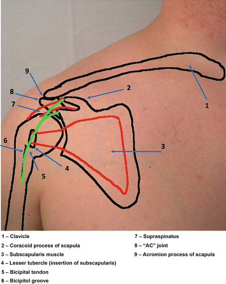
Fig. 3.2
Surface anatomy of the shoulder – anterior view
The shoulder is actually composed of four joints: the sternoclavicular (SC) joint, the acromioclavicular (AC) joint, the glenohumeral (GH) joint, and the sternothoracic (ST) joint (see Fig. 3.3). Pathology can occur at any one of these joints, but pathology is most common in the AC and GH joints. The labrum provides some static stability to the GH joint, as does the joint capsule, which is composed of three main ligaments: the anterior, inferior, and posterior glenohumeral ligaments. Injury to these ligaments can allow the humerus to slide out of the glenoid fossa. When this occurs to a minor degree and spontaneously relocates, this is called subluxation; if the humeral head completely leaves the socket, it is true dislocation. Many children and adults have some degree of physiologic subluxation due to natural laxity of these ligaments and do not necessarily have underlying pathology [3].
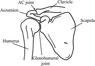
Fig. 3.3
Skeletal anatomy of the shoulder – anterior view
The muscles provide dynamic support to the shoulder joint. The biceps tendon crosses this joint and provides additional dynamic support, but the majority of stability is due to the deltoid muscle, which applies constant upward force on the shoulder, and the muscles of the rotator cuff (supraspinatus, infraspinatus, teres minor, and subscapularis), pulling the “ball into the socket,” opposing the deltoid’s anterior pull. Due to their location, the supraspinatus and infraspinatus tendons are the most common muscles injured in the rotator cuff [4].
Red Flags
Age of Patient. A very elderly or very young patient complaining of shoulder pain may represent a more serious type of condition. This would include pathological fracture, growth plate injury, and malignancy.
Nonmusculoskeletal Causes of Pain. Symptoms such as shortness of breath, GI upset, cough, rash, weight loss, fever, multiple joint involvement, or morning stiffness should prompt the investigation of metabolic causes of shoulder pain.
Trauma. Patient’s trauma to the shoulder should receive a radiographical evaluation to rule out fracture. Additionally, those with major trauma can have visceral pain that is referred to the shoulder (such as lung or chest wall injury or ruptured spleen) and should be carefully evaluated.
Suspected or Known Dislocation. These patients should have an X-ray to rule out fracture. Posterior shoulder dislocation can be difficult to identify. In patients with a history of seizure disorder or electrocution, consideration for posterior dislocation should be entertained.
General Approach to the Patient
There has been a great deal written about the physical examination of the shoulder. Many physical examination techniques have been described to detect various pathologies of the shoulder. The accuracy of these examination techniques has been called into question. Recent meta-analysis of the available medical literature has not shown a high correlation between physical exam findings and final diagnoses discovered either on MRI or in surgery [5–8]. Shoulder complaints are common, comprising up to 16 % of all musculoskeletal visits to any healthcare provider [9, 10].
The clinician should approach all shoulder complaints with a history and physical examination. Detailed testing can be performed depending on suspected conditions described below.
A basic history should include duration, nature, and location of pain as well as inquiring about any radicular symptoms. Risk factors for nonmusculoskeletal conditions that can present with shoulder pain (such as lung disease, MI, etc.) should be assessed. Patients who appear unstable after trauma or who are suspected to have life-threatening nonmusculoskeletal pathology (such as MI or concern for ruptured viscus) should not be assessed in the primary care outpatient setting and should be transported to an emergency department.
Examination should start with complete exposure of the shoulder, with either the shirt removed or the patient wearing a sleeveless shirt or sports bra. A gown can be tied around the trunk under the arm, if needed for modesty. The skin should be evaluated for any rashes or lesions that might suggest other causes of pain, such as shingles [9].
A brief neurovascular exam is important for ALL patients who present with shoulder pain. The Spurling’s maneuver (described in Chap. 2) can be quickly performed and if negative rules down radiculopathy. Hoffman’s test (flicking middle finger with resulting flexion of index finger and thumb) may point to an upper motor lesion causing the patient’s discomfort. Evaluation of the radial pulse and capillary refill is reassuring for adequate distal circulation.
Palpate the SC and AC Joints. Tenderness over the SC joint usually occurs only after significant direct trauma to the anterior chest and is a cause for concern, as trauma this severe can sometimes be associated with traumatic shoulder dislocations. Palpate the AC joint for tenderness.
Next, have the patient abduct the arm from the side to the overhead position, noting pain or limitations of ROM. From behind the patient, evaluate the scapula for winging or abnormal movements with shoulder abduction, which may indicate damage to the long thoracic nerve [10].
Common Clinical Presentations
Rotator Cuff Pathology
With age, most patients get some degree of external impingement of the tendons of the rotator cuffs, causing rotator cuff tendinopathy. These patients will complain of generalized anterolateral shoulder pain that may radiate toward the deltoid. Examination of these patients involves impingement testing, which can be accomplished with Hawkins and Neer testing (shown in Fig. 3.4). Negative findings on both these tests have an LR− of 0.1 for impingement, nearly ruling out this pathology [5].
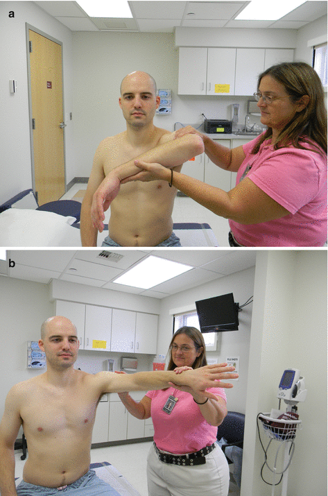
Fig. 3.4
Hawkins (a) and Neer (b) testing
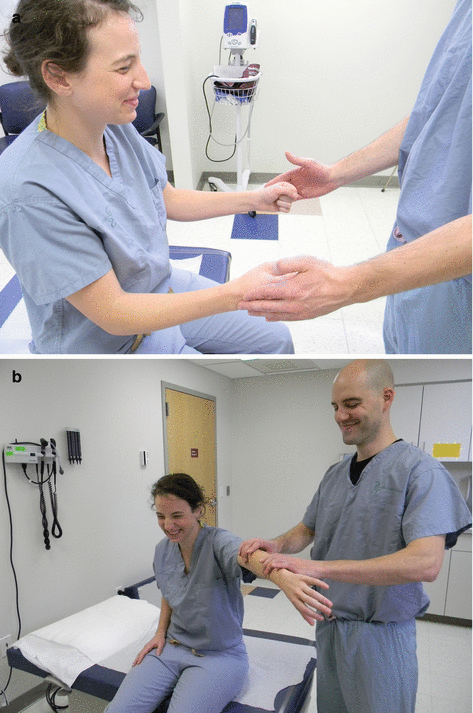
Continued impingement or a traumatic event can lead to rotator cuff injury, in which the tendon is completely torn. In addition to the impingement testing, the supraspinatus and infraspinatus strength should be tested. Supraspinatus weakness is tested by having the patient extend the elbows, abduct the arms to 90°, and forward flex about 45°. The patient then makes a fist with “thumbs down” and then resists the examiner putting downward pressure on the arms. Infraspinatus weakness is tested by having the patient keep the elbows at the sides with the elbow flexed 90°. The patient then pushes out (externally rotating) against resistance from the examiner. The supra- and infraspinatus tests are depicted in Fig. 3.5. It is important to understand that an abnormal test result is one that demonstrates true weakness of the movement, not just lack of effort due to pain [4]. These three tests together (impingement testing, supraspinatus weakness, and infraspinatus weakness) can be very useful in predicting rotator cuff tears. If no abnormal results are present, a tear is basically ruled out. Presence of only one is not predictive, but the presence of two of these has an LR+ of 5 for tear, and the presence of all three has an LR+ of 48 for rotator cuff tear [5, 6].