CHAPTER 14 The Role of Arthroscopy in Midcarpal Instability
Introduction
The concept of midcarpal instability (MCI) has evolved slowly since it was first described in 1934. Many investigators have contributed to our understanding of this condition over the years, which led to the consolidated classification by the senior author (DML) (Table 14.1).1 It appears that MCI represents several distinct clinical entities differing in the cause and direction of subluxation but sharing the common characteristic of abnormal force transmission at the midcarpal joint. The following discussion centers on intrinsic MCI. Extrinsic MCI due to a dorsally mal-united distal radius fracture is treated by a distal radius osteotomy and hence falls outside the scope of this discussion.
Table 14.1 Classification of Midcarpal Instability
| Intrinsic | Extrinsic |
|---|---|
| A. Palmar | A. Distal radius mal-union |
| B. Dorsal | |
| C. Combined |
Pathomechanics
The mechanism of the clunk in palmar midcarpal instability (PMCI) has been described in detail by Lichtman et al.2 The palmar arcuate ligament complex is comprised of a radial arm that is confluent with and distal to the radioscaphocapitate (RSC) ligament and an ulnar arm or the triquetrohamate-capitate ligament (TCL) (Figure 14.1). In the normal situation, the proximal carpal row moves smoothly from a flexed position when the wrist is in radial deviation to extension when the wrist is in ulnar deviation. This is due to the progressive tightening effect of the arcuate ligament as it stretches out to length (which incrementally pulls the midcarpal row into extension) and to the carpal bone geometry, which causes the triquetrum to translate dorsally along the helicoidal facet of the hamate. When the arcuate ligament is attenuated, this synchronous motion is lost.
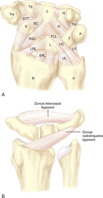
Studies by Trumble et al.3 and Viegas and coauthors4 have shown that sectioning either the TCL or the dorsal radiocarpal (i.e., dorsal radiotriquetral ligament) can produce a volar intercalated segmental instability (VISI) deformity and simulate PMCI. More recently, Lichtman showed in vivo that tightening the DRCL alone can stabilize the proximal carpal row and eliminate the clunk of PMCI—emphasizing the potential importance of dorsal ligament laxity in the pathogenesis of this disorder.5 The senior author now believes that PMCI is caused by laxity of both the TCL and the DRCL, which allows an excessive palmar sag of the heads of the capitate and hamate at the midcarpal joint. This produces a VISI pattern of the proximal row in the nonstressed wrist. This sag results in a loss of joint contact across the midcarpal joint, which manifests clinically as a loss of the smooth transition of the proximal row from flexion to extension as the wrist deviates ulnarward.
The proximal carpal row thus stays in a flexed position until the terminal extent of ulnar deviation, when the helicoidal shape of the hamate facet suddenly forces the triquetrum dorsally. This snaps the lunate and subsequently the scaphoid into extension, causing a sudden reversal of the VISI. This sudden proximal row extension is responsible for the painful and rapid catch-up clunk that occurs. As the wrist moves back to neutral, the triquetrum translates down the hamate facet—which allows the proximal row to drop back into VISI while the distal row again settles palmarly into its slightly subluxated starting point (Figure 14.2a and b).
The dorsal pattern of MCI has not been studied as extensively. It appears that laxity of the radial arm of the palmar arcuate ligament permits the capitate and hamate to translate dorsally to an excessive degree, especially with ulnar deviation of the wrist.6,7 It is of note that in both the palmar and dorsal patterns the proximal row always moves into extension and the distal row translates dorsally with ulnar deviation. It is the timing and force of this movement that differentiate the two patterns.
Diagnosis
Clinical Findings
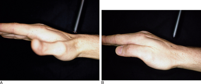
In PMCI the patient presents with a history of clunking of the wrist. Patients can often reproduce the clunk on both sides because generalized ligamentous laxity frequently coexists. However, the patient may have a trivial injury that accentuates this normal laxity—resulting in a painful clunk. Upon physical examination, close inspection will reveal a sag of the midcarpal joint with the wrist in radial deviation—which is reduced with active or passive ulnar deviation (Figure 14.3a and b). The clunk may be reproduced by performing the midcarpal shift test.2 This test is performed by placing the patient’s wrist in neutral with the forearm in pronation. A palmar force is then applied to the hand at the level of the distal capitate. The wrist is simultaneously loaded and deviated ulnarly. The test result is positive if a painful clunk occurs that reproduces the patient’s symptoms.
Related posts:


Stay updated, free articles. Join our Telegram channel

Full access? Get Clinical Tree


