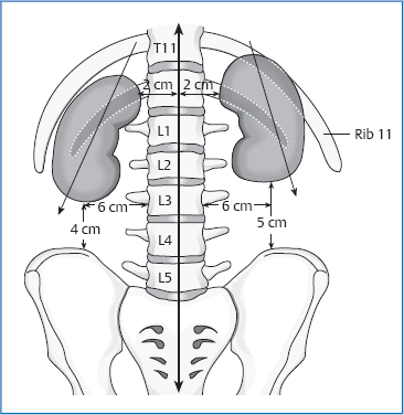
14 The Kidneys Size: 12cm long, 7cm wide, and 3cm thick. Posterior Left kidney: Upper pole: T11 Renal pelvis: L1 Lower pole: L3 The right kidney is located approximately 1–1.5cm lower than the left kidney. Fig. 14.1 Location of the kidneys. Anterior Left kidney: Upper pole: rib 9 Lower pole: 1–2cm above the navel Right kidney: Upper pole: rib 9 Lower pole: level of the navel The axis of the kidney runs slightly diagonally from cranial–medial to caudal–lateral. This consists of an anterior leaf and a posterior leaf. Both leaves merge superior and lateral to the kidneys. This “fascial sac” is open on the bottom. The fascias of both kidneys merge at the level T12–L1 in front of the spinal column. Retrorenal lamina: This covers quadratus lumborum and psoas major and is fixed anteriorly and laterally to the spinal column (medial to the psoas and diaphragm). Prerenal lamina: This lies next to the peritoneum and Toldt fascia. On the left side, it is associated with this fascia in a larger area. It covers the kidney, hilum, and the large prevertebral vessels. Both laminae surround the adrenal glands, merge superiorly, and are attached to the diaphragm. Inside the fascial layers and surrounding the kidney, we find fat (fat capsule). This exists from about age 10 on. Fig. 14.2 Connections of the right kidney. Posterior Anterior Right kidney: Left kidney: The adrenal glands lie superior to both kidneys. Fig. 14.3 Connections of the left kidney. Renal artery (originates in the aorta, roughly 1 cm below the superior mesenteric artery; the left one is shorter than the right one). Renal vein (left vein is longer than the right one, ends in the inferior vena cava). Maximal time: 5–7p.m. Minimal time: 5–7a.m. For basic information, see page 34. Three factors determine the movement of the kidneys: The engine of this movement is the diaphragm. During inhalation (20000/day, 600m/day), the kidney moves 3–4cm caudally. The upper pole is pressed forward during inhalation (psoas slide rail). In addition, the kidney moves in a caudal–lateral direction and rotates outward. During inhalation, we feel a movement from medial–cranial to lateral–caudal in connection with an outward rotation (“windshield wiper”). During exhalation, the kidney completes the opposite movement. Definition. Urinary stones in the kidney and excretory urinary tracts. Causes. Excessive amounts of stone-forming substances in the urine. Risk factors include: Clinical. Asymptomatic if the calculi do not constrict the urinary tracts. Obstructing stone causes: Definition. Infection of the upper urinary tract caused by pathogenic organisms. Causes. Highly virulent organisms coinciding with a weakened state of defense. Precipitating factors include: Clinical Definition. Complex of symptoms, consisting of: Causes. We find primary or secondary preexisting glomerular disorders, e.g.: Clinical Definition. Most common form of malignant tumor in the kidney, in most cases originating in the tubular cells. Causes. Degeneration of proximal tubular cells.
Anatomy
General Facts
Location
Renal Fascia
Topographic Relationships
 diaphragm and psoas arcade
 pleura (indirectly in the area of the costodiaphragmatic recess up to the level of L1)
 rib 12, on the left also rib 11
 psoas major and its fascia
 quadratus lumborum and transversus abdominis
 subcostal, iliohypogastric, ilioinguinal nerves
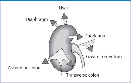
 liver
liver
 hepatoduodenal ligament
 right colic flexure
 transverse mesocolon
 duodenum, descending part
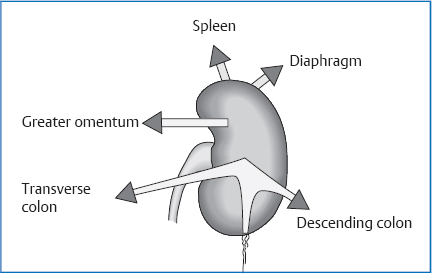
 spleen
 stomach
 pancreas
 duodenojejunal flexure
 jejunum
 left colic flexure (stronger fixation than on the right)
Attachments/Suspensions
 turgor
 pressure of other organs and tonicity of the abdominal muscles
 fat capsule
 hilar vessels and ureter (braking function)
 thoracic suction effect and tonicity of the abdominal muscles during respiration
Circulation
Arterial
Venous
Lymph Drainage
 lumbar nodes
 lumbar trunk
 thoracic duct
Innervation
 sympathetic nervous system from T10 to L1 via the lesser and lowest splanchnic nerves and the lumbar splanchnic nerves 1 and 2 to the celiac plexus, aorticorenal ganglion, renal plexus, and posterior renal ganglion
 vagus nerves (via the celiac plexus)
 sacral parasympathetic part (S2–S4) via the superior hypogastric plexus to the renal plexus
Organ Clock
Organ–Tooth Interrelationship
Movement Physiology according to Barral
Mobility
Motility
Physiology
Functions of the Kidney
 regulation of the fluids and electrolytes
 regulation of the acid–base balance
 excretion of substances through the urine (urea, creatinine, uric acid, etc.)
 excretion of foreign substances (medications)
 regulation of blood pressure (renin–angiotensin– aldosterone system)
 hormone production (erythropoietin, renin, calcitriol, prostaglandins)
 degradation of peptide hormones
Pathologies
Symptoms that Require Medical Clarification
Nephrolithiasis
 lack of physical movement
 insufficient fluid supply
 familial predisposition
 medications (calcium, vitamin C and D therapy)
 gout
gout
 diabetes mellitus
 kidney disorders
 hyperparathyroidism
 colic with hematuria
 nausea
 vomiting
 abdominal pain
 flank pain
 pain radiating into the genitals and inside of the thighs
Acute Pyelonephritis
 stricture of the urinary tract
 vesicoureteral reflux
 neurogenic disturbance of bladder voiding
 calculi
 diabetes mellitus
 immunosuppressive therapy
 pain elicited by percussion in the kidney area
 flank pain
 headache
 sweating
 nausea
 vomiting
 fever >38.5°C
Nephrotic Syndrome
 proteinuria
 hypoproteinemia
 dysproteinemia
 hyperlipoproteinemia
 edemas
 poststreptococcal glomerulonephritis
 rapidly progressive glomerulonephritis
 systemic disorders, e.g., lupus erythematosus
 microhematuria
 edemas
 hypertonicity
Renal Cell Carcinoma
Clinical
 hematuria
 elevated ESR
 palpable abdominal mass
 hypertonicity
 weight loss
 anemia
 intermittent fever
 asymptomatic in the early stages
Osteopathic Practice
Cardinal Symptoms
Related posts:


Stay updated, free articles. Join our Telegram channel

Full access? Get Clinical Tree


