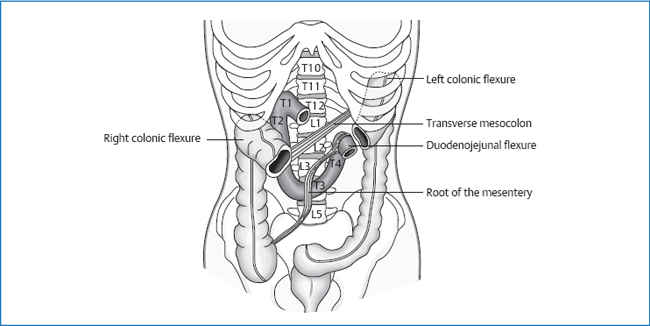
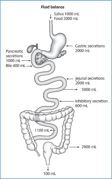
12 The Jejunum and Ileum The total length of the jejunum/ileum is 5–6 m, of which two-fifths is the jejunum and three-fifths the ileum. This section of the small intestine starts at the duodenojejunal flexure and ends at the ileocecal valve, where it runs into the cecum. Arrangement of the small intestine is into 15–16 loops, the jejunum more horizontal and the ileum more vertical. Furthermore, the jejunum is located more around the navel, while the ileum is found in the right lower abdomen. As a whole, the jejunum and ileum lie more on the left side: the loops cover the descending colon while the ascending colon remains exposed. The root of the mesentery is approximately 12–15 cm long and 18 mm wide. It extends from the duodenojejunal flexure to the ileocecal valve and so crosses L2–L5 in a diagonal course. Fig. 12.1 Location of the root of the mesentery. At the level of L3–L4, the superior mesenteric vein enters the mesentery. Between L4 and L5, the root crosses the ureter on the right side. The mesoappendix originates in the mesentery and continues into the appendico-ovarian ligament. At its distal end, the root crosses the testicular/ovarian vein. Superior mesenteric artery. Portal vein. Along the vessels to the superior mesenteric lymph nodes—celiac and lumbar lymph nodes. Maximal time: 1–3p.m. Minimal time: 1–3 a.m. For basic information, see page 34. The diaphragm has only a minor effect on this area of the small intestine. Nevertheless, we can deduce from the type of suspension that large movements must be taking place in the jejunum and ileum. These are the intrinsic movements necessary for mixing the food mash and propelling the chyme (peristalsis). In the expiratory phase, the entire bundle of the small intestine performs a clockwise rotation; during the inspiratory phase, it goes back in the opposite direction. In these sections of the intestine, we find the typical wall structure of the entire digestive tract (esophagus to rectum). The layers are: The inner surface of the intestine feels smooth like velvet. This is the result of the 0.5–1-mm high fingerlike projections into the mucosa (villi), which are arranged close to each other in evenly spaced intervals. At the base of these villi, we find the tubular phlegm-producing glands of the intestines (crypts) which sink deeply from this base into the depth. The epithelial cells of the villi are furthermore marked by evaginations in the membrane (ciliated border, microvilli), which increase the surface of the intestine, as well as the villi and crypts, by many times: the inner surface of the intestine thereby reaches almost twice the size of the surface of the skin (4 m2). The villi are the site of absorption, and the crypts the site of regeneration and secretion. Diverse food particles are absorbed by the greatly enlarged surface of the villi, whereas the crypt cells provide for the renewal of dead epithelial cells and of the mucus that covers the inner surface of the intestine. The tela submucosa consists of connective tissue. It includes: Mucosa and submucosa form the circular crossfolds of the intestine (valves of Kerckring = plicae circulares), which are visible with the naked eye. They serve to enlarge the surface area and decrease in number distally. The muscular layer consists of smooth muscle cells that are arranged in an inner ring-shaped and an outer longitudinal muscle layer. The peristaltic and chyme-mixing movements originate in these muscles. In a layer of connective tissue between these two layers, we find the Auerbach plexus (myenteric plexus), which supplies the two muscular layers vegetatively. The adventitia is a layer of connective tissue that is very pronounced in those areas of the intestine that are not covered by peritoneum. In the area of the jejunum and ileum, this layer is therefore only very thin and called subserosa. This is the visceral peritoneum. Jejunum. In the proximal parts of the jejunum, the valves of Kerckring and villi are very dense, and the ciliated border contains a particularly large quantity of enzymes: most of the absorptive processes for carbohydrates, fats, and proteins occur in the first 100 cm of the jejunum. Distally, the valves of Kerckring decrease in number and height, but we find more lymphatic follicles. Ileum. Distally, the valves of Kerckring disappear completely; instead, we find a large number of Peyer patches, which are involved in immune defense. These two sections of the intestine are the main location for the digestion and absorption of fats, carbohydrates, proteins, vitamins, inorganic salts, and water. The digestive enzymes produced by the small intestine are located partly in the ciliated border on the lumen side; others are spread diffusely in the cytoplasm of the epithelial cells and released only after the cells die off. The short lifespan of the cells in the intestinal mucosa, i.e., 2–3 days, accommodates this physiology. Each day, the body absorbs 8–9 L of water with 50–100g electrolytes in the small intestine, but of these only 1.5 L comes from food; the rest is discharged as digestive secretions from the intestine. The α-amylase in the saliva and pancreas breaks starch down into oligosaccharides. Together with disaccharides from food, these are further broken down by enzymes in the ciliated border into monosaccharides and in this form absorbed by the membrane. With the assistance of bile salts, lipases in the saliva and pancreas split triglycerides in the food into monoglycerides and free fatty acids. In combination with the products of fat digestion and fat-soluble vitamins, the bile salts form micelles. Micelles attach themselves to the epithelium of the small intestine and mediate the absorption of the products of fat digestion into the mucous membrane. The bile salts themselves are entered into an enterohepatic cycle in the terminal ileum. Fig. 12.2 Sites of absorption for individual food components. The acid gastric juice denatures the proteins in the food—it dismantles the three-dimensional structure of proteins. Pepsins in the gastric juice then split the proteins into medium-long and short peptides. The pancreatic enzymes (trypsin and chymotrypsin) further cleave the proteins into oligopeptides, which are then broken down by enzymes in the ciliated border into amino acids, or di- or tripeptides, and absorbed by the mucosa in the small intestine.
Anatomy
General Facts
Location
Root of the Mesentery

Topographic Relationships
Anterior and Cranial
 transverse colon
 transverse mesocolon
 greater omentum
 anterior abdominal wall
Posterior
 posterior parietal peritoneum
 kidneys
 ureter
 aorta
aorta
 inferior vena cava
 common iliac vein
 duodenum
 descending and ascending colon
Caudal
 bladder
 uterus
 rectum
Lateral
 ascending colon
 abdominal wall
 cecum
cecum
 sigmoid colon
Attachments/Suspensions
 organ pressure
 turgor
 root of the mesentery
Circulation
Arterial
Venous
Lymph Drainage
Innervation
 sympathetic nervous system from T10 to T12 via the minor splanchnic nerve to the superior mesenteric ganglion
 vagus nerve
Organ Clock
Organ–Tooth Interrelationship
Movement Physiology according io Barral
Mobility
Motility
Physiology
Microscopic Wall Structure
 mucous membrane
 tela submucosa
 muscular layer
 adventitia
 serosa
Mucous Membrane (Mucosa)
 epithelium
 lamina propria mucosae (reticular connective tissue)
 lamina muscularis mucosae (smooth muscle)
Tela Submucosa
 the Meissner plexus (submucous plexus), which supplies the smooth muscle and the glands
 circulation for the mucosa
 the Peyer patches (lymph follicles that increase in number distally)
Muscular Layer
Adventitia
Serosa
Regional Differences in Wall Structure between the Jejunum and Ileum
Processes of Absorption in the Jejunum and Ileum
Digestion of Carbohydrates
Digestion of Fats

Digestion of Proteins
Musculoskeletal Key
Fastest Musculoskeletal Insight Engine


