Fig. 8.1
Skeletal anatomy of the hip – anterior view
Red Flags
Systemic symptoms, such as fever or weight loss, may indicate systemic illness, such as infection (septic joint) or malignancy, and these patients should be evaluated thoroughly to rule out major pathology.
Pain that is worse at night is also concerning for malignancy.
Patients who are completely unable to bear weight should also be evaluated immediately by X-ray. Inability to bear weight may indicate fracture or avascular necrosis of the femoral head [1].
Approach to the Patient with Hip Pain
As with any chief complaint, it is important to obtain a thorough history. History should include onset, location, and duration of pain, as well as risk factors including past medical history, past surgical history, history of injury or trauma, and medication use. Activity level including regular exercise, or lack thereof, and occupational activities and exposures should also be included. History is very helpful in determining the etiology of the pain and will guide your focused clinical exam. The location of the pain can be very helpful in organizing your differential diagnosis, as the causes of anterior, posterior, and lateral hip pain are often very different. Also, a patient complaining of “hip pain” may actually be experiencing symptoms stemming from the low back or pelvis, so these should remain on a working differential diagnosis.
Hip Exam
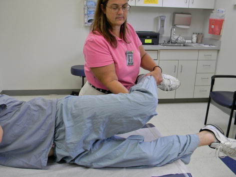
All hip exams should include the following: gait assessment; assessment of range of motion (ROM) of the hip, as well as brief neurovascular exam, strength testing, including distal pulses, and straight leg raise and/or slump test, to rule out radicular etiology. Limited ROM, particularly of internal rotation, suggests intra-articular pathology, such as a fracture, or arthritis. Normal external rotation ranges from 30 to 45°, internal rotation from 20 to 35°, and flexion from 120 to 125° [1].
Anterior Hip Pain
In the adult population (pediatric patients are discussed in Chap. 12), the most concerning etiologies of anterior hip pain are those involving the joint itself, or intra-articular causes. These include osteoarthritis (OA) and femoroacetabular impingement (FAI).
OA of the hip occurs typically in patients over the age of 50. In addition to age, other risk factors such as obesity, prior injury, and family history may predispose a patient to OA. Elements of the history that may indicate OA are as follows: groin pain that is worse with activity, morning stiffness usually lasting less than 30 min, and specific activity triggers, such as getting into and out of a car. On physical exam, pain with active or passive internal or external rotation (particularly internal), often, is an indication of intra-articular pathology such as OA. Radiographs are not necessary to make the diagnosis but should be obtained if other diagnoses are being entertained or conservative therapy has failed. Treatment involves weight loss, analgesic medication, PT, steroid injection, and ultimately hip replacement, when all other treatment measures fail to control or manage the symptoms [1].
Femoroacetabular impingement (FAI) is the abnormal articulation of the acetabular rim and the proximal femur. It can lead to injuries of the labrum and articular cartilage and can lead to osteoarthritis of the hip if left untreated. Elements of history that may indicate FAI include the following: anterolateral hip pain, aggravated by prolonged sitting, leaning forward, or pivoting motions in the young adult population [2]. On physical exam, range of motion will often reproduce the pain for patients, as will many special maneuvers. The most sensitive of these is the FADIR test or flexion, adduction, and internal rotation (Fig. 8.2) [2].


