Fig. 5.1
Surface anatomy of the hand – palmar aspect

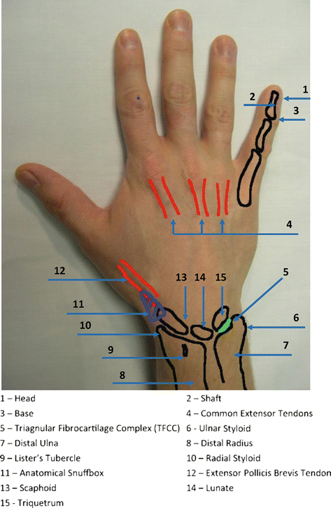
Fig. 5.2
Surface anatomy of the hand – dorsal aspect

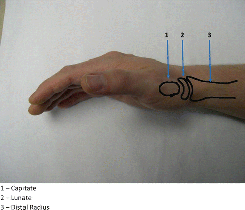
Fig. 5.3
Surface anatomy of the wrist– lateral view

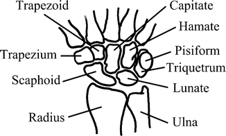
Fig. 5.4
Skeletal anatomy of the wrist – palmar aspect
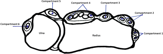
Each of the digits has two neurovascular bundles, one on the radial side and the other on the ulnar side, which contain an artery, vein, and nerve [3]. The extensor tendons, which originate on the lateral dorsal forearm, insert on the dorsal hand. The flexor tendons from the medial forearm insert on the palm of the wrist and hand [4]. The superficial flexor tendon on each phalynx inserts at the base of the middle phalynx, while the deep flexor tendon inserts on the base of the distal phalynx. Figure 5.5 and Table 5.1 demonstrate the extensor and flexor tendons of the fingers.


Fig. 5.5
Flexor and extensor tendons
Table 5.1
Flexor and extensor tendons
Compartment | Structure in compartment | Common pathologies | Clinical findings |
|---|---|---|---|
1 | Extensor pollicis brevis | DeQuervain’s tenosynovitis | Fibrosis of the synovium around the first dorsal compartment; pain, crepitus radial aspect of the wrist |
Abductor pollicis longus | |||
2 | Extensor carpi radialis brevis | Intersection syndrome | Fibrosis of intersection between the first and second dorsal compartment, approximately 4 cm proximal from Lister’s tubercle on the radial aspect of the wrist |
Extensor carpi radialis longus | |||
3 | Extensor pollicis longus | Rupture with distal radius fracture | Degenerative tear of the extensor pollicis longus tendon |
Drummer boy palsy | Patient cannot extend the thumb | ||
4 | Extensor digitorum communis | Tendinopathy | Tendinopathy and pain on dorsal aspect of the wrist with wrist extensor |
Extensor indicis proprius | |||
Posterior interosseous nerve | |||
5 | Extensor digitorum minimi | Vaughn–Jackson syndrome | Rupture of the extensor digitorum minimus and sometimes the extensor carpi ulnar. Patient has weakness extending the index finger and small finger |
6 | Extensor carpi ulnaris | Snapping wrist | Extensor carpi ulnaris tears or subluxation on ulnar when ulnar retinaculum is damaged. Causes painful snapping on ulnar aspect of wrist |
The metacarpal–phalangeal (MCP) joint of the thumb differs from the usual “ball and socket” joints of the other digits. Instead, it is a “saddle” joint, which allows for the pincer grip. This joint is largely supported by soft tissue and is therefore easily injured.
Red Flags
Several hand and wrist conditions should be urgently investigated or referred due to potential serious sequelae.
Compound fracture. Any compound fracture should be urgently referred to a specialist. Active or profuse bleeding should be controlled with pressure; no attempts at exploration should be made.
“Fight bite.” A fight bite occurs when the fist strikes a tooth of another person, usually over the knuckles of the ring or little fingers. This type of injury carries high risk of penetration of tendon or even bone, and even a very small mark on the skin may overlie a more serious injury. These injuries are at high risk for infection and should be referred for surgical exploration.
Burns. Severe burns, especially on the palmar side of the hand or wrist, carry risk of underlying tendon injury and should be referred for management.
Injury from high–pressure tools. Air and paint guns can cause high-pressure injury to underlying structures with only a small entry point in the skin and should be referred.
Stay updated, free articles. Join our Telegram channel

Full access? Get Clinical Tree


