Fig. 4.1
(a) Absolute growth of elderly population in the United States, subdivided by age group. From 2000 to 2050, the population of persons over the age of 65 in the United States is expected to increase from 35 million to 87 million (Source of data: United States Census Bureau). (b) Growth of proportion of total US population classified as elderly 2000–2050. The population of patients over age 85 is the fastest growing age group in the United States (Source of data: United States Census Bureau)
Not only are the elderly becoming a larger part of the population, but they are often involved in trauma, which is the fifth leading cause of death in that age group [4]. Patients over the age of 65 represent 12 % of the population but 28 % of all fatal injuries in the United States [5]. In addition, they sustain a disproportionate number of fractures [6]. The lifetime prevalence of hip fractures is 1 in 3 in women and 1 in 12 in men [7]. In a study of a Medicare database, it was determined that between 1986 and 2005, the annual mean number of hip fractures was 957 per 100,000 in females and 414 per 100,000 for males [8]. One problem encountered in treating elderly trauma patients is that most traditional treatment protocols are designed for younger patients. The elderly differ in important ways from young and middle-aged adults. When caring for them, providers should take into account their special characteristics, such as increased risk of mortality, physiologic changes, and preexisting medical conditions/comorbidities [9].
Mortality
The most important factor for the orthopaedic surgeon to consider when caring for elderly patients is their increased risk of mortality compared to younger age-cohorts. Increased mortality in elderly patients has been correlated with higher Injury Severity Score (ISS), lower Glasgow Coma Scale (GCS) score, as well as greater transfusion and fluid resuscitation requirements (Table 4.1) [10]. One review of 100 trauma patients aged ≥65 compared with 100 younger controls found that geriatric trauma patients were six times as likely to die as their younger counterparts despite having similar ISS scores. Mortality in the group aged 65 and older was 17 % compared to 3 % in controls [11]. The presence of shock was found to increase mortality significantly among older patients [11]. Hospital and the intensive care unit (ICU) length of stay as well as costs were also higher in the elderly group. The mechanisms of injury in the geriatric patients were more likely to be a simple fall or pedestrian-car injury versus motor vehicle accidents (MVA), gunshot wounds, or crush injuries for younger patients [11]. Similar findings have been reported found in other studies, including one that demonstrated 42 % mortality in patients > [greater than] 65 years of age compared to 20 % in younger patients admitted to a level one trauma center. The same study also reported a mortality rate of nearly 50 % in patients >75 years of age [9].
Table 4.1
Factors associated with increased mortality in geriatric trauma patients
Higher ISS |
Lower GCS |
Greater transfusion and fluid resuscitation requirements |
More advanced age |
Increased number of preexisting medical conditions |
In a review of 7,798 trauma patients, the effect of comorbidities was studied. Despite having similar ISS and GCS, those with preexisting comorbidities had higher mortality; 9.2 % mortality was seen in those with comorbidities compared to 3.2 % in those without [12]. Mortality increased with the number of preexisting diseases: 15.5 % in those with ≥2 and 24.9 % in those with ≥3 comorbidities. The highest mortality was seen in those with renal disease, malignancy, and cardiac disease [12]. In a review of the Pennsylvania Trauma Systems Foundation database of 33,781 geriatric patients, an overall mortality rate of 7.6 % was reported [13]. The investigators also found that, for each 1 year increase in age beyond age 65, the odds of dying after geriatric trauma increased by 6.8 % [13]. Furthermore, the same investigators demonstrated that when controlling for vital signs, GCS, and ISS, comorbidities (hepatic disease, renal disease, cancer, congestive heart failure, chronic obstructive pulmonary disease, and chronic steroid in descending order of association with mortality) had a significant effect on mortality on these older patients [13]. Notably, warfarin use had no effect on odds of death. Preexisting medical conditions increase complications and contribute to both early and late mortality in the geriatric trauma patient [2].
Clement et al. demonstrated that early mortality is correlated with more severe injuries (higher ISS). Late mortality, occurring more than 13 days after injury seen in patients with lower ISS, was most often due to medical complications [14]. Patients over the age of 65 and those with ISS less than 16 were at higher risk for late mortality: 33.3 % versus 12 % in younger patients with similar injury scores [14].
Increased mortality is also seen when looking at specific injuries in the elderly. In a review of 234 pelvic fractures treated at a single trauma center, investigators found that older patients were 2.8 times more likely to receive blood and required more blood transfusions (7.5 vs. 5.0 units) than younger pelvic trauma patients [15]. Lateral compression fractures (LC) occurred 4.6 times more frequently than anterior-posterior compression (APC) fractures in the elderly. In addition, the lateral compression fractures were minor in older patients (98 % were grades LC 1 or 2), but these patients were four times more likely to require blood. Overall, older patients had a higher mortality rate even after adjusting for ISS [15].
Most published data on mortality after geriatric fractures pertains to hip fractures. The mortality after hip fractures that are surgically treated is 9 % at 30 days, 19 % at 90 days, and 30 % at 12 months [16]. Other studies have demonstrated a 20 % or greater mortality within 1 year of sustaining a hip fracture in geriatric patients [17, 18]. The risk of mortality is highest in the first year after the fracture [19]. This increase in risk of mortality decreases over the first 2 years but never returns to the baseline rate [20]. Surgical treatment of hip fractures is associated with a 4 % mortality risk [21]. Age at the time of fracture is known to be a significant risk factor. In one prospective series of 1,109 patients with hip fracture, mortality risk was found to increase 4 % for each additional year of age [22].
In those over age 60, mortality is also increased after other major types of osteoporotic fractures, including those of the vertebrae, pelvis, distal femur, multiple ribs, and proximal humerus [23]. Even seemingly benign injuries can have significant effects on geriatric patients. One study found that, in this population, each additional rib fracture increases the risk of pneumonia by 27 % and the risk of mortality by 19 % [24]. It is likely that deaths from traumatic injury are underreported as these patients often die from complications that are recorded as the cause of death instead of the true cause, trauma [6].
Initial Triage
Considering the increased mortality associated with trauma in older patients, extra vigilance should be maintained when treating them, even prior to arrival at the hospital. Under-triage is common and harmful to geriatric patients. A review of the Florida trauma system found that under-triage of patients older than 55 years of age occurs 71 % of the time [25]. When triaging patients in the field, paramedics and emergency medical technicians used a trauma scorecard consisting of physiologic criteria (systolic blood pressure less than 90 mmHg, respiratory rate less than 10 or greater than 29 bpm, GCS less than 12) as well as anatomic and mechanistic criteria (second- and third-degree burns greater than 15 % body surface area, paralysis, ejection from vehicle, amputation proximate to wrist or ankle, penetrating injury to the head, neck, chest, abdomen, or groin) to determine whether a patient should be classified as a trauma alert, which occurred if any of the eight criteria were met. Patients classified in this way were taken to a trauma center for immediate care by a dedicated trauma team. This under-triage is thought to occur because older patients often do not exhibit hypotension or tachycardia in response to a significant trauma.
When initially evaluating geriatric trauma patients, it is important to recognize that normal presenting vital signs may not accurately reflect injury severity [2, 26]. There are two reasons for this situation. Polypharmacy can significantly alter the elderly patient’s response to injury: beta blockers, for example, can mask hypotension and tachycardia [27]. The second reason is the frequent presence of comorbidities. Preexisting hypertension in geriatric patients should be considered. A normal blood pressure for a younger adult may represent hypotension in the older geriatric patient [1].
Another potential reason for under-triage is the mechanism of injury in the elderly. Triage criteria often fail to identify cases of major trauma from falls [1]. Treating physicians may also fail to recognize the significant trauma that may result from a fall in a geriatric patient. A review of 26,025 patients from a single metropolitan area found being elderly or female to be two of the most significant risk factors associated with being under-triaged [28].
Because of high under-triage rates, some centers have added old age as a criterion for trauma team activation. In an effort to avoid missing patients who might benefit from activation of the trauma system, Demetriades et al. suggested trauma team activation for all patients more than 70 years old [29]. In a follow-up study, the same group created a new protocol for trauma team activation: age less than 70 years, systolic blood pressure less than 90 mmHg, heart rate less than 120 bpm, respiratory rate less than 10 or greater than 29 bpm, unresponsive, or emergency department physician judgment. In addition to the new trauma activation protocol, the patients received early invasive monitoring and resuscitation, as well as early ICU admission. The authors studied 336 patients with ISS >15 and found a decrease in mortality from 53.8 to 34.2 % [30]. Another group uses patient age >55 as a criterion for considering transportation to a trauma center [3]. Further support for the move to consider age as an indication to activate a trauma team in addition to standard hemodynamic and mechanistic criteria is research which has shown that older patients still have a relatively high risk of death even in the absence of physiologic abnormality and should thus be treated more aggressively [9, 26]. Further research in this area is needed to establish the inclusion of advanced age as a criterion for trauma team activation.
The sentiment that trauma centers have significantly better outcomes than community care hospitals in treating older injured patients has been borne out in the literature. In a retrospective review of the very elderly (≥80), Meldon et al. found that patients taken to level II trauma centers experienced less mortality (5.2 %) than those taken to community care hospitals (9.9 %) [3]. Mortality at level I centers was higher (24 %), but these hospitals also cared for patients who were injured more seriously than the other groups. For very old patients, location of treatment for geriatric trauma patients is an important factor.
Mechanism of Injury
One of the largest and most comprehensive studies examining the mechanisms and outcomes of serious injury in geriatric trauma patients was performed by Richmond et al. [31]. They queried the Pennsylvania Trauma Systems Foundation database for seriously injured patients and excluded those who sustained isolated hip fractures after falls from standing height. A total of 38,707 patients over the age of 65 years over a 10-year period were included in the study. Mechanisms of injury were, in descending order of rank, as follows: falls (61.7 %), motor vehicle collision (22.6 %), others (9.4 %), pedestrian (4.6 %), hit by object (0.9 %), and assault (0.8 %) (Fig. 4.2) [31]. As patients get older, falls become the most common mechanism, responsible for 49.2 % of traumas in the 65–74 age group and 81.1 % in the above 85 age group. Extremities and the pelvic girdle were the most injured body regions in 47.4 % of cases [31]. Another multicenter study found the cause of injury in patients ≥65 years of age to be falls (40.6 %), motor vehicle collision (20.2 %), pedestrian struck (10 %), others (7 %), gunshot wound (5.5 %), stab wound (2.6 %), motorcycle collision (0.4 %), and unknown (0.3 %) (Table 4.2) [32]. Physicians should also be cognizant of elder abuse, which has an estimated prevalence of 32 cases per 1,000 persons [33]. Unlike child abuse, there are no fracture patterns that are considered pathognomonic for elder abuse. It should be suspected when there are ambiguous, inconsistent stories that do not match with the presenting injury.
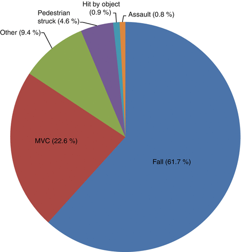
Fig. 4.2
Mechanisms of injury in trauma patients older than 65 years of age. Falls and motor vehicle collisions (MVC) are the most common mechanisms of traumatic injury in elderly patients, accounting for more than 80 % of trauma cases in one series [31]
Table 4.2
Relative frequency and fatality rate of various mechanisms of traumatic injury in geriatric patients [32]
Mechanism | Relative frequency (%) | Fatality rate (%) |
|---|---|---|
Fall | 40.6 | 11.7 |
MVC | 28.2 | 20.7 |
Pedestrian struck | 10.0 | 32.6 |
Other | 7.0 | 13.8 |
GSW | 5.5 | 52.1 |
Stab wound | 2.6 | 17.3 |
MCC | 0.4 | 11.8 |
Unknown | 0.3 | 19.0 |
Even though falls from a standing height often are relatively benign in other population groups, they are significant in the elderly, as they can cause multiple injuries which may result in an ICU admission similar to a higher-energy mechanism. Older adults can sustain multiple injuries from low-energy trauma [34]. The reason for the increased incidence of falls in the elderly is multifactorial. Physiologic changes take place with aging, including decreased visual, auditory, proprioceptive, and vestibular inputs, which combine with diminished reaction times, unsteady gait, and loss of strength and coordination to contribute to the increased likelihood of falls. This may be compounded by cardiac dysrhythmias and orthostatic hypotension of various etiologies, including polypharmacy [1].
Finally, it is important not only to think about the mechanism of injury but also the reason that the patient became injured. An older individual injured in a motor vehicle collision may have had a transient ischemic attack, stroke, or arrhythmia. Other causes to consider include hypoglycemia or dementia (which interferes with the ability to recognize and avoid road hazards). The underlying reason could be more benign, such as presbyacusis, presbyopia, or slowed reaction time. The cause of the trauma can not only significantly alter the patient’s immediately course of care but could also prevent future injuries.
In summary, there are a number of possible mechanisms for injury in a geriatric patient with falls being the most common. However, because the older segment of the population is staying active, an increasing number of injuries from all mechanisms are expected [2].
Physiology
When considering physiology, there are several important differences between old and young adults. Older adults have reduced physiologic reserves and diminished compensatory mechanisms and are therefore less able to deal with the added stress presented by trauma. This narrow physiologic tolerance and the restricted reserves should be expected when managing geriatric trauma patients [1]. A patient’s chronologic age may not equal their physiologic age, which is modulated by their preexisting conditions (Table 4.3). What makes management of these patients difficult is that their comorbidities may be unknown at the time of presentation.
Table 4.3
Common comorbidities, or preexisting conditions, that are frequently encountered in geriatric patients
Cardiovascular | Coronary artery disease, congestive heart failure, hypertension, peripheral vascular disease |
Pulmonary | Shunting, chronic obstructive pulmonary disease |
Renal | Decreased renal function |
Gastrointestinal | Malnutrition |
Central nervous system | Cerebral atrophy, dementia, cervical stenosis |
Dermatologic | Thinner subcutaneous tissue |
Endocrine | Diabetes mellitus |
Musculoskeletal | Osteoporosis, previous orthopaedic hardware, or implants |
Aging of the cardiovascular and respiratory systems reduces the older patient’s ability to respond to hypoxia and shock [1]. The heart of a geriatric patient, often affected by coronary artery disease and congestive heart failure, accommodates poorly to hypovolemia. A low cardiac rate aggravates decreased preload from hypovolemia, resulting in decreased cardiac output. This in turn results in myocardial ischemia and an additional drop in cardiac output. Older patients have increases in resting ventilation/perfusion mismatch resulting in pulmonary shunting, which make them more susceptible to developing hypoxia. Many also have chronic pulmonary obstructive disease, which can complicate their perioperative management.
Aging is accompanied by diminished renal function and decreased creatinine clearance, which is masked by a serum creatinine that is falsely normal because their muscle mass is lost. It is not uncommon for older patients to have some degree of malnutrition, which can impair healing and recovery. Their dura mater becomes adherent to the cranium, eliminating the epidural space [1]. With concomitant age-related brain atrophy, the bridging veins are more susceptible to injury. Consequently, epidural bleeds are less frequent but subdural bleeds are more common in the older population. Preexisting cervical stenosis places the patient at significant risk for central cord syndrome. Their integument has impaired thermoregulation and loss of subcutaneous cushion, the latter of which can lead to more degloving injuries. Both diabetes and peripheral vascular disease complicate wound healing and predispose to infection and nonunion [6]
The musculoskeletal system, affected by muscle atrophy and osteoporosis, is often subject to more severe injuries even when less kinetic energy is imparted on the limb. When a previous orthopaedic implant is present such as a joint replacement or other implant, it almost always alters the fracture pattern and treatment for a given injury. The effect of osteoporosis in the care of the geriatric patient cannot be overemphasized.
Osteoporosis
Osteoporosis is a condition of decreased bone mineral density resulting from an imbalance of bone formation and resorption. Osteoporosis is divided into primary and secondary osteoporosis. Primary osteoporosis is the loss of bone mass associated with the aging process, since peak bone density is attained in young adulthood and decreases steadily thereafter. Secondary osteoporosis is due to a variety of causes, including insufficient intake of calcium or vitamin D, gastrointestinal malabsorption, metabolic derangements (hyperparathyroidism, hypo- or hyperthyroidism, Cushing’s syndrome, renal pathology), medications (anticonvulsant drugs, prednisone), and deficiencies of gonadal hormone (estrogen deficiency, low testosterone levels) (Table 4.4) [35]. The majority of these causes can be detected with laboratory tests [35]. Recommended tests include a basic metabolic panel with serum calcium levels, a 24-h urine calcium measurement, a 25-hydroxy-vitamin D level, as well as thyroid-stimulating hormone and parathyroid hormone levels. Other tests can be ordered as indicated based on history and physical examination. In addition, all patients with fragility fractures should be considered for dual-energy X-ray absorptiometry (DXA) scans and prescriptions for calcium and vitamin D supplements [35]. Bukata et al. stated that, when caring for patients with geriatric fractures, the identification of potentially correctable etiologies of impaired bone quality should be sought [35]. Failure to do so may impair recovery and places the patient at increased risk for future fractures.
Table 4.4
Common causes of secondary osteoporosis that should be considered in evaluating patients with fragility fractures [35]
Insufficient intake of calcium |
Insufficient intake of vitamin D |
Gastrointestinal malabsorption |
Hyperparathyroidism |
Hypothyroidism |
Hyperthyroidism |
Cushing’s syndrome |
Renal pathology |
Medications (anticonvulsant drugs, prednisone) |
Estrogen or testosterone deficiency |
Osteoporosis is common, affecting 45 % of women and 15 % of men over the age of 50 [36, 37]. The lifetime prevalence of osteoporosis is 13–18 % in women and 3–6 % in men [38]. In postmenopausal women with fragility fractures, the prevalence is 30 % [39]. This number is higher for lower-risk patients such as men and premenopausal women [40]. Known risk factors for osteoporosis include female gender, multiparity, BMI < [less than] 18.5 kg/m2, smoking, excessive alcohol consumption, certain medications, and northern European or Asian ancestry.
Since the architecture of the bone is compromised by osteoporosis, fractures in osteoporotic bone may display a high-energy pattern and be challenging to treat in spite of being a low-energy mechanism [35]. In addition, the altered mechanics of osteoporotic bone make fixation difficult. Failure of hardware in osteoporotic bone typically occurs at the bone-implant interface, resulting in cutout, fracture subsidence, or pull off of the plate. Failure happens when the load transmitted at the bone-implant interface exceeds the diminished strain tolerance of osteoporotic bone [37]. Based on a cadaveric study on pullout strength in human tibiae whose bone density was assessed with CT scanning, Seebeck et al. demonstrated that decreased cortical thickness and loss of trabecular bone make it difficult to obtain good purchase with standard hardware in osteoporotic patients [41]. Consequently, constructs that maximize surface contact area between the hardware and the bone are preferred [35]. Examples include hardware with locking screws, screws with larger diameter, and bicortical screw purchase. Load-bearing, as opposed to load sharing, devices are preferred in these patients [35]. In addition to proper implant selection, the treating surgeon should perform thorough preoperative planning and accurate fracture reduction when treating these patients.
A number of studies have shown that orthopaedic surgeons have not been proactive in identifying patients who may benefit from treatment of their osteoporosis. In a study of 1,162 older female patients who sustained distal radius fractures, only 24 % of patients underwent either diagnostic evaluation or treatment for osteoporosis. In addition, those who were older were significantly less likely to be treated appropriately with antiresorptive agents [36]. A similar study was performed to assess management of osteoporosis in older women who sustained low-energy femoral neck fractures and found calcium supplements and antiresorptive medications to be under-prescribed at the time of discharge from the hospital [38]. A similar, more recent study found a 20 % treatment rate with osteoporosis in older patients who sustained distal radius fractures [40]. Orthopaedic surgeons are often the first to see the patient with fragility fractures. This pattern, combined with the fact that treatment options exist that are effective in reducing the rate of additional fractures, provides an additional reason for the orthopaedist to initiate the evaluation for osteoporosis or metabolic bone disorders and either commence treatment or refer the patient to a provider who can do so [6]. It has been suggested to assess and treat patients with fragility fractures or provide referral for osteoporosis care. One center reported a >95 % rate in successful diagnosis, treatment, or referral after implementation of such a program, which involved a team consisting of a dedicated coordinator supported by surgeons, residents, allied health-care professionals, and administrative staff [42]. Administration of bisphosphonates, which are first-line agents in the treatment of osteoporosis, is important because they have been shown to decrease fracture rates [6].
Secondary Fracture Prevention
In addition to selecting the appropriate procedure to perform, the orthopaedic surgeon should also start patients on calcium and vitamin D supplementation to correct any preexisting vitamin D deficiency and optimize fracture healing in osteoporotic patients. In a study of 954 patients at metabolic bone clinics, 73–89 % were found to have levels of vitamin D below the normal range (32 ng/mL or 80 nmol/L). In those with hip fractures, this figure was 84–96 % [43]. When vitamin D levels fall below 10 ng/mL (25 nmol/L), the patient is at risk for secondary hyperparathyroidism, which can further complicate bone metabolism [44]. There is mounting evidence that all patients with fragility fractures should have their vitamin D levels normalized, but current recommendations are under review [35]. One group of authors made the recommendation that orthopaedists should correct 25-hydroxy-vitamin D levels to more than 32 ng/mL, as this is the level at which PTH secretion normalizes [45].
There are two forms of vitamin D supplementation: ergocalciferol (vitamin D2), which is derived from plant and yeast sources, and cholecalciferol (vitamin D3), which is derived from animal sources and produced in the skin [35]. Numerous protocols exist for vitamin D supplementation. Bukata et al. suggest administering 50,000 IU ergocalciferol weekly for a duration that depends on baseline vitamin D levels: over 5–8 weeks if 20–30 ng/mL, 16 doses if 10–19 ng/mL, and 24 doses if less than 10 ng/mL [35]. This is followed by daily cholecalciferol (2,000 IU) in addition to vitamin D contained in their multivitamin or calcium supplements as long-term therapy [35]. With normalized vitamin D levels and bone metabolism optimized, the patient is at a reduced risk for fragility fractures.
Prevention of falls represents the other aspect of secondary prevention of fractures. Falls cause 90 % of fractures of the forearm, hip, and pelvis in geriatric patients [6]. Risk factors for falling include the use of sedatives (benzodiazepines, phenothiazines, antidepressants), cognitive or visual impairment, lower extremity disability, foot problems, balance or gait abnormalities, neurologic conditions, and use of an assistive device for ambulation [46, 47]. Strategies to reduce the risk of falls include ensuring that the patient has vision correction, proper shoe wear, discontinuation of excessively sedating medications, and modification of the home environment [48]. Examples of the latter include providing good lighting throughout the home, lowering beds, installing carpet over hard floors, eliminating throw rugs and thick carpets, and providing grab bars in the bathroom [49]. Unfortunately, even with the best efforts at prevention, fragility fractures still occur. When they present to the hospital emergency department, there are several steps that can be taken to provide care that is both appropriate and expedient.
Interdisciplinary Comanagement
Advances in medical and anesthetic management have permitted less healthy geriatric patients to undergo orthopaedic surgical procedures successfully that may have been contraindicated in the past because of preexisting conditions [6]. When interviewing these patients, it is important to determine not just their past medical histories but to inquire about which medications they are currently taking, including antihypertensives and anticoagulants. Beta blockers should not be discontinued, as this can trigger rebound hypertension. The management of anticoagulants should be discussed with the consulting medicine provider. Reversal of warfarin (Coumadin) should be done with oral vitamin K or fresh-frozen plasma or while waiting for hepatic synthesis of clotting factors; the international normalized ratio (INR) should be ≤ [less than or equal to] 1.5 before the patient is taken to the OR [35]. Patients on clopidogrel (Plavix) or other platelet inhibitors should not receive spinal or epidural anesthesia but may undergo early surgery under general anesthesia [35]. Many patients who are on clopidogrel after placement of drug-eluting stents are at increased risk for stent thrombosis if they discontinue the drug. Therefore, risks and benefits of operating on a patient under the effect of clopidogrel must be balanced with platelet transfusions. Similar considerations should be made with respect to newer anticoagulants such as dabigatran (Pradaxa) and rivaroxaban (Xarelto), as these medications have no direct reversal agents and take 1–2 days to be eliminated from the body. Medications to avoid include nonsteroidal anti-inflammatory drugs (NSAIDs, as they impair bone healing and kidney function) centrally acting antihistamines, meperidine, most antiemetics, benzodiazepines, H2 (histamine) receptor antagonists, and anticholinergics [35, 50].
Most patients benefit from medical evaluation and optimization prior to surgery, as doing so will ultimately lead to decreased time to surgery and length of stay [50, 51]. Most have comorbidities and are at increased risk for adverse outcomes and postoperative complications. Comorbidities increase the risk of functional decline and death [19, 50]. Geriatricians are trained to address these comorbidities and manage potentially adverse outcomes and complications, thereby helping to maximize outcomes [50]. The medicine specialists should coordinate overall medical care of these older patients, both preoperatively and postoperatively [50].
The comanagement model of orthopaedics and geriatrics, characterized by the comanagement of the patient by geriatricians and orthopaedists who share responsibility throughout the patient’s hospital stay, was developed in England in the 1950s [50, 52]. It has been shown to reduce complications, length of stay, readmission rate, cost of care, and mortality, as well as lead to better function, higher levels of patient, and provider satisfaction [50]. In-hospital mortality rates for comanaged hip fractures have been shown to range from 0.6 to 11 % [50]. Under this model, each of the two specialists sees the patient, writes his or her own orders, and communicates with the other specialist on a daily basis (Fig. 4.3, Table 4.5) [50]. Frequent communication between medical and surgical providers reinforces the rationale between treatment decisions for the patient and provides continuous education for the providers [50]. An interdisciplinary team of health-care professionals should provide support for the patient and the admitting physicians [52].
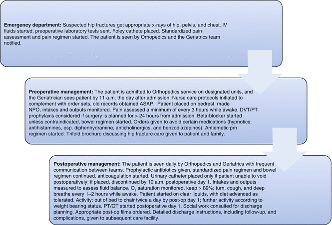
Fig. 4.3
Standardized protocol for comanagement of hip fractures patients in the Geriatric Fracture Center at Highland Hospital, University of Rochester Medical Center, Rochester, NY [50]
1. Most patients will benefit from surgical management of their fracture |
2. The shorter the delay to surgery, the less time to develop iatrogenic complications |
3. Comanagement with frequent communication avoids iatrogenesis |
4. Standardized protocols decrease unwarranted variability in patient care |
5. Discharge planning begins immediately, at the time of admission |
In general, this model has resulted in reduced short-term mortality and complications and increased 1-year survival compared to traditional models where only orthopaedic surgeons are responsible for the management of the patient [52]. However, implementing such a program requires considerable effort on the part of the physicians as well as administrative support and strong leadership for continuous monitoring and improvement of the model once it is implemented. Additional studies are needed to evaluate the model’s cost-effectiveness and long-term outcomes as well as its applicability to lower volume hospitals. It is believed that approximately 100 cases per year are needed to develop sufficient expertise in managing these patients [53]. The limited number of geriatricians combined with good availability of hospitalists implies that hospitalist comanagement will become an important variation of the geriatric comanagement model.
Another point regarding consultation is important—the need for a cardiology consultation. This decision should be made by a geriatrician or hospitalist. In the case of a complex cardiac condition, these consultations represent a common reason for delay of fixation of fractures. Some authors feel that these consultations are usually unnecessary in patients with geriatric fractures [35].
Other salient points on perioperative management of geriatric fracture patients are worth mentioning. Dehydration is almost always present on admission, so immediate hydration with normal saline and administration of red blood cells as indicated preoperatively will help minimize the risk of hypotension upon induction of anesthesia [35]. Polypharmacy is common in these patients. Harmful or unneeded medications should be discontinued by the consulting medical provider while the patient is admitted [35].
Familiarity with geriatric patients on the anesthesiologist’s part is important to ensure safe and efficient anesthetic care. The American Society of Anesthesiologists classification of surgical risk is useful to assess preoperative risk secondary to comorbidities [6]. Special considerations that anesthesiologists must take into account when caring for geriatric patients is that they are at significant risk for aspiration pneumonia and they do not tolerate excessive hypotension. Most orthopaedic procedures may be performed with either regional or general anesthesia. Studies have shown no difference in short-term or long-term mortality or functional treatment in hip fractures performed under spinal or general anesthesia [54, 55]. Efficacy of regional anesthesia (spinal/epidural) as prophylaxis against deep vein thrombosis and pulmonary embolism has been previously demonstrated [56].
An important principle in preoperative management of geriatric fractures is to provide the patient with a prompt work-up so as to minimize delays in surgery. This minimizes the length of bed rest for the patient, which is associated with venous thromboembolism, skin breakdown, pulmonary decompensation, delirium, and infection [50]. Additionally, surgical delays have been shown to affect the mortality. Delays of more than 48 h have been shown to increase 30-day mortality by 17 % in an analysis of 18,209 Medicare patients treated for hip fractures [57]. In a published model of geriatric fracture care, the authors suggest that surgical cases should be treated as urgent but not emergent and should only proceed to the operating room once the patient is optimized for surgery [50]. In a subsequent publication by the same group, the average time from admission to the operating room was <24 h [19]. Another way to expedite care is admission directly from a nursing home or assisted-living center to the hospital floor. This can eliminate delays in the emergency department and may reduce time to surgery [50]. Other authors have found that having a standardized pathway for geriatric fractures that are either directly admitted or present to the emergency department leads to a shorter length of stay, lower mortality, both in-hospital and 1-year mortality [58].
Surgical Management Strategies
When determining how a patient’s orthopaedic injuries are to be treated, it is important to obtain a detailed history of his or her functional status, including what if any assistive devices are used for ambulation at baseline, how independent the patient is with activities of daily living, where the patient lives, with whom, how many floors he or she has they have in his or her their residence and how many steps to enter, and whether a first-floor living situation can be arranged. The patient’s medical and cognitive histories are also important because the information contained in these histories has an impact on surgical options and potential for rehabilitation [6]. Given the multitude of factors and complexity of decision-making related to patient care, treatment recommendations should be made by the surgeon after an informed discussion with the patient, his or her family members, and other consulting providers. These are summarized in Table 4.6 and described in more detail later in this section as well as in their respective chapters throughout the rest of this text.
Fracture location | Character | Options | Comments |
|---|---|---|---|
Femoral neck | Nondisplaced | Three cannulated screws | Allow impaction with weight-bearing, stabilizing the fracture, expect some shortening to occur |
Sliding hip screw with antirotation pin | |||
Displaced | Hemi or total arthroplasty | Physiologically younger, active patients without dementia may qualify for total hip arthroplasty | |
Physiologically older, low-demand patients and all dementia patients receive hip hemiarthroplasty | |||
Controversy as to whether cemented or press-fit stems are better; limited evidence that cemented stems are a slightly better choice | |||
Pertrochanteric | Stable | Sliding hip screw | Maintain tip-apex distance of less than 27 mm to prevent cutout |
Unstablea | Trochanteric entry nail | Position implant in the center of femoral head to avoid failure of fixation | |
Use long nail for subtrochanteric fractures | |||
Plating associated with increased rate of mechanical failure and nonunion | |||
Distal femur | 95° condylar blade plate | Inexpensive but unforgiving implant | |
Distal femoral locking plate | Can be placed percutaneously, expensive, requires intraoperative fluoroscopy | ||
Retrograde intramedullary nailing | Limited distal fixation, can be used in periprosthetic fractures if the implant has an open box, expensive, requires intraoperative fluoroscopy | ||
Proximal humerus | Simple, minimally displaced | Nonoperative management | Satisfactory outcomes in most cases but radiographs less appealing |
Simple, displaced | Locked plate | Lack of medial buttress can result in varus collapse and screw penetration of humeral head, requires intraoperative fluoroscopy, high complication rate | |
3 and 4 part
Related posts:Stay updated, free articles. Join our Telegram channel
Full access? Get Clinical Tree
 Get Clinical Tree app for offline access

|


