Fig. 6.1
Testing the superficial and deep flexor tendons. (a) Test the superficial flexor tendon by immobilizing the base of the finger and have the patient flex the PIP joint. (b) Test the profundus or deep flexor tendon by immobilizing the finger just distal to the PIP joint and have the patient flex the DIP
Red Flags
1.
High-Pressure Injury. If the patient is working with any power equipment with air or liquid substance under pressure, this injury can occur. The patient may only complain of a stinging or burning sensation. A very small puncture wound may be observed, but no discernable wound sometimes occurs with these injuries. These injuries require urgent consultation. Even if left untreated for a few hours, a great deal of damage can occur to the affected finger.
2.
Bites. Any type of bite requires special care. In the wrist chapter, there was discussion on the “fight bite.” Bites may occur from any mammalian, marina, or insect to the finger or hand. Upon close inspection, one should determine if there is any retained foreign body. Table 6.1 (Daniels 2004) reviews the evaluation and treatment of uncomplicated puncture wounds from animal bites.
Table 6.1
Evaluation and treatment of uncomplicated puncture wounds
Clinical situation | Antibiotic prophylaxis |
|---|---|
Low-risk, traumatic injuries (clean wounds with easily demarcated borders, no devitalized tissue) | None |
Injuries in immunocompromised patients (e.g., patients with human immunodeficiency virus infection, diabetes) | Gram-positive cocci coverage |
Wounds with devitalized tissue | Gram-positive cocci coverage if wound tendon or joint space is contaminateda |
Animal and human bites (other than superficial abrasions) | First-generation cephalosporin. In patients with bites that may contain Pasteurella multocida or Eikenella corrodens, consider penicillin or amoxicillin-clavulanate potassium (Augmentin). In immunocompromised patients, consider erythromycin or amoxicillin-clavulanate. In patients with sepsis and petechial rash, consider intravenous ciprofloxacin (Cipro) and clindamycin (Cleocin)b |
Puncture wounds | Case-by-case decision |
3.
Tendon Injuries. Figure 6.2 shows the appearance of a lacerated flexor or extensor tendon. The profundus flexor tendon (deep tendon) which attaches to the base of the distal phalanx may be avulsed when a patient’s hand or finger is forced into extension. The common term for this is called “jersey finger,” and it occurs most often on the ring and small finger. Commonly, the mechanism of injury occurs when a player grabs a jersey during a football game and the finger is partially extended when the opposing player runs away. These conditions must be identified and treated quickly to avoid major dysfunction of that phalanx.
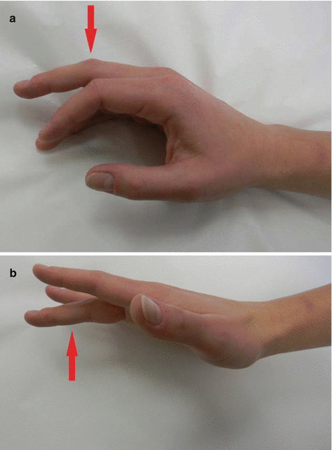
Fig. 6.2
Testing for lacerated flexor or extensor tendon. (a) Lacerated flexor tendon. (b) Lacerated extensor tendon
4.
Unstable Fractures. Any open fracture is a medical emergency, and prompt consultation of a hand surgeon is necessary. Many fractures with benign appearance on radiograph can oftentimes be unstable. A general rule of thumb is if over one-third of the joint is involved with the fracture, it is unstable. Radiographs of any traumatized phalanx should be obtained as some fractures can cause malalignment of the fingers. Figure 6.3 reviews how this can easily be checked.
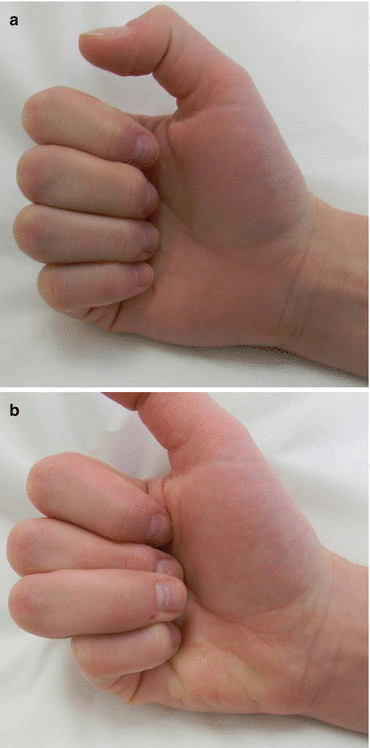
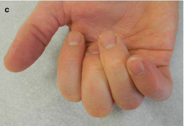
Fig. 6.3
Malalignment of fingers. (a) Demonstrates the patient flexing their fingers and there is no scissoring. (b, c) Demonstrates how the patient’s fingers will scissor or crossover when they flex
5.
Septic Tenosynovitis. The flexor tendon has a sheath that can be punctured and become infected. The sheath becomes inflamed and fills up with purulent material. Kanavel’s cardinal signs occur. This includes slight digital flexion, uniform volar swelling, flexor tendon sheath tenderness, and pain on extension. It is not necessary for all four of these signs to be present to make the diagnosis. Purulent tenosynovitis should highly be suspected with increased swelling after puncture wound or on the volar surface of the hand.
6.
Get Clinical Tree app for offline access

Acute Trauma. The area on the palmar surface of the hand between the MP joint and the carpal bones has been referred to in the past as “no man’s land.” There are a large number of intrinsic and extrinsic tendons in this area that can be damaged. Any patient with a deep laceration to this part of the hand should have a surgical consultation. Finger amputation or maceration is also a medical emergency. Table 6.2 reviews the indications/contraindications for reattachment of an amputated digit. The residual finger should be gently cleansed and irrigated with saline and wrapped in nonadherent petroleum gauze and bulky dressing put in place. The amputated digit should be wrapped in nonadherent gauze, moistened with saline, and put into a sterile container or tied-off plastic bag. The amputated part should not be manipulated or submerged in water. The plastic bag or container should be placed in a larger container with ice. No tissue of any type should be removed or debrided prior to consulting the replant surgeon.
Table 6.2


Indications and contraindications for attempted reattachment of an amputated finger or hand
Related posts:


Stay updated, free articles. Join our Telegram channel
Full access? Get Clinical Tree