Chapter 2 The diagnosis of fractures and principles of treatment
How to diagnose a fracture
1 History
Diagnosis
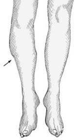
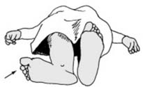
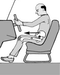
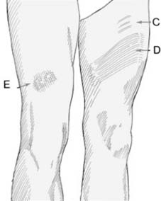
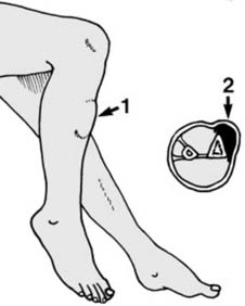
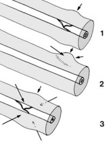
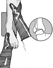
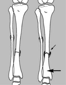
2 Inspection (a) Begin by inspecting the limb most carefully, comparing one side with the other. Look for any asymmetry of contour, suggesting an underlying fracture which has displaced or angled.
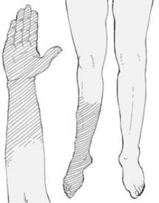
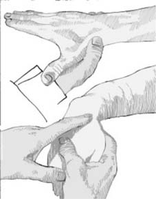
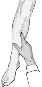
8 Inspection (g) Note the colour of the injured limb, and compare it with the other. Slight cyanosis is suggestive of poor peripheral circulation; more marked cyanosis, venous obstruction; and whiteness, disturbance of the arterial supply. Feel the limb, and note the temperature at different levels, again comparing the sides. Check the pulses, and the rapidity of pinking-up after tissue compression.
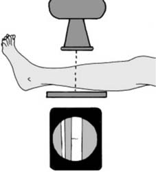
17 Localised views Where there is marked local tenderness, but routine films are normal, coned-down localised views may give sufficient gain in detail to reveal for example a hair-line fracture: if such films are also negative, the radiographs should be repeated after an interval of 10–14 days if the symptoms are persisting (see also Hair-line Fractures in Ch. 1/Frame 13)
Other visualisation techniques
1 CT (CT) scans
4 MRI scans
These avoid any exposure to X-radiation and produce image cuts as in CT scans with a greater ability to distinguish between different soft-tissues. In the trauma field they are of particular value in assessing neurological structures within the skull and spinal canal, and meniscal and ligamentous structures about the knee and shoulder.
Pitfalls
Treatment of fractures
Primary aims
The primary aims of fracture treatment are:
Initial management
A = Airway
B = Breathing
C = Circulation
Classification of haemorrhage
Estimating blood loss
The following list gives a crude guidance in anticipating potential blood loss:
Further resuscitation and assessment
Fluid replacement:
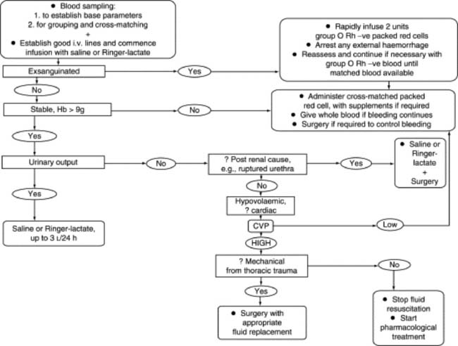
The volume of the replacement required can vary enormously, and therefore must be judged by the response: see the following flow chart (Fig. 2.1) for a summary of replacement management.
Assessing the response to replacement:
There is varying opinion on the best methods of assessing the stability of the circulation and the success of resuscitation. In all, the degree and maintenance of a positive response to treatment is more important than the reading of isolated values. Many methods are advocated including the following:
Stay updated, free articles. Join our Telegram channel

Full access? Get Clinical Tree


