I. INTRODUCTION AND PHILOSOPHY
A. Epidemiology of orthopaedic trauma. Musculoskeletal trauma has gained increased public attention over the past 15 years for a number of reasons. Such reasons include the realization of societal impact from health care costs and lost workdays in the labor force. These statistics are coupled with an increased understanding that the orthopaedic surgeon and health care team can positively influence outcomes, both through intervention and education on injury prevention. Leadership from key organizations (Orthopaedic Trauma Association, Arbeitsgemeinshaft fur Osteosynthesisfragen, American Academy of Orthopaedic Surgeons, American College of Surgeons [ACS], American Orthopaedic Association, and others) has played a major role in lobbying for proactive trauma-related health care policy and implementation of public education programs for injury prevention.
Although it is likely that educational measures such as seat belt safety, aggressive standards for highway safety, and lower blood alcohol limits for drivers help to lower accident-related injury rates, other forces seem to counter such progress, such as the pervasive trend toward faster cars and the burgeoning enthusiasm for extreme sports and the use of cell phones and texting while driving. In addition, new and significant musculoskeletal injuries are being seen in areas such as those due to airbags in survivors of motor vehicle accidents.
An even greater awareness is emerging regarding the aging baby boomers who will account for massive demands on the healthcare system. The baby boomers will be hitting the 65-year-old age mark in 2011, and it is estimated that by the year 2040 there will be 35,000,000 more people over the age of 55 than there are now and that the number of hip fractures alone will increase from 250,000 to 500,000 on an annual basis.1 Geriatric musculoskeletal trauma is due to the vulnerability of the skeletal system from the natural process of relative bone mineral loss manifesting in the condition of osteoporosis. Compounding the number of injuries in this group is the increasingly active lifestyle of this aging population and increasing lifespan. To put it in perspective, it is estimated that one-third of all women reaching the age of 90 will sustain at least one hip fracture.2
B. Definition of musculoskeletal trauma. Musculoskeletal trauma includes any injury to bone, joint (including ligaments), or muscle (including tendons). Nearly always, such injuries occur in combination, as the energy imparted to breaking a bone or tearing a ligament is also dissipated to impact structures nearby or even distant from the most obvious site. With greater experience, such combinations of injuries become more apparent to the diagnostician, which leads to swifter and more accurate detection of injury characterization.
C. Multiple injuries. The energy it takes to render trauma to the musculoskeletal system can also injure other organs. This is particularly common with the high-energy mechanisms that are responsible for pelvic, spine, shoulder girdle, or long bone fractures. Owing to the greater density and strength of bone in younger individuals, there are even greater energy required to create fractures in this population. Therefore, it is incumbent upon the trauma team to remain vigilant to the likelihood of injuries to other bones and other organ systems. Often, the dramatic and salient injuries, so-called distracting injuries, during the initial patient evaluation will attract the diagnostic and therapeutic attention, whereas occult and sometimes equally grave injuries remain initially undetected.
For example, it is estimated that only 7% of the patients who die from life-threatening high-energy pelvic fractures actually die from arterial exsanguination related to the pelvic fracture itself,3 whereas the rest succumb due to injury involving other organ systems, with head injury representing the predominant cause.4 Forty percent of patients with femur fractures have other associated fractures,5 and 90% of patients with scapula fractures have other associated injuries.6 Heightened awareness when evaluating the trauma patient will keep the missed injury rate to a minimum.
D. Missed injury rate. The missed injury rate in the context of polytrauma has been reported to be 4% to 18%.7 It is important to drive down this rate with appropriate protocols that underscore the importance of the secondary survey, as well as a re-review of the patient’s physical examination each ensuing day after injury. A secondary survey is a head-to-toe review by a physician that occurs after the initial primary survey, which is defined as the evaluation of the patient’s airway, breathing, circulation, disability, and exposure, followed by screening imaging of the cervical spine (spiral computed tomography [CT] scan), anteroposterior chest, and pelvic X-ray).
The main reasons cited for missing injuries include multisystem trauma with another more apparent orthopaedic injury, trauma victim being too unstable for a full orthopaedic evaluation, altered sensorium, hastily applied initial splints obscuring injuries, and inadequate radiographs.8
E. Multiple patients. It is not uncommon, particularly at a Level I trauma center, to require simultaneous evaluation of multiple patients, such as with motor vehicle collisions in which multiple victims are involved. Doctors who have had some training on the fundamentals of trauma surgery and, in particular, Advanced Trauma Life Support (ATLS), which includes strategies for triaging patients and resources during a mass casualty situation, must be available in order to effectively “captain the ship.” ATLS courses have been developed, refined, and sponsored by the ACS and have an excellent educational track record. Typically (but not exclusively), in the United States, it is a general surgery trauma surgeon who is running the trauma room. It is beyond the scope of this orthopaedic text to delve into the specifics of ATLS management; however, we will focus on some of the fundamentals and cover the triage process of multiple orthopaedic injuries that may present during such circumstances. To further master the details of ATLS management, please refer to the ATLS Manual (8th edition) published in 2008.
It is imperative to understand what is an orthopaedic emergency and what is orthopaedically urgent. A review of the “orthopaedic emergencies” in a subsequent section of this chapter will help to understand how these injuries need to be prioritized for treatment. Furthermore, it is important to understand what measures can be taken to staged orthopaedic treatment. Not all broken bones need immediate definitive treatment, and the practitioner must understand how to titrate the proposed treatment to the physiologic presentation of the patient. For example, a patient with limited physiologic reserve, due to a great physiologic challenge from hemorrhagic shock and compromised ventilation from a hemothorax, should not spend excessive hours in the operating room getting several fractured bones stabilized. In such a case, it may be prudent to place an external fixator across a broken femur rather than to immediately nail the femur or place a plaster splint on a displaced ankle fracture rather than to fix it right away. These measures save a lot of time, blood loss, anesthesia, fluid challenge, and avoid the “second hit” during a potentially critical stage in postinjury physiologic evolution.
There are many ways to stage the treatment of injuries, which also gives the orthopaedist more time to solicit expertise, get to know the patient and family, plan the details of an operation, and understand the comorbidities and the likelihood of patient compliance. All these different factors may, in fact, impact the ultimate treatment that the orthopaedic surgeon chooses to render and will most certainly influence positive outcomes.
II. EVALUATING THE TRAUMA PATIENT FROM THE ORTHOPAEDIC PERSPECTIVE. The patient who presents from an accident scene should receive a much different type of workup than would be called for by a scheduled history and physical examination, although certainly history and physical examination are part and parcel of the process.
It is important to acknowledge that there is a “captain of the ship,” typically an ATLS-trained general surgeon, who will have the clearest overview of the patient and who will be delegating many simultaneous responsibilities. The person taking primary responsibility for the orthopaedic injuries must heed the captain’s call and clearly communicate diagnostic or treatment priorities for the orthopaedic conditions and ultimately fit those into the context of overall priorities.
Trauma care is organized in three stages: primary survey, secondary survey, and definitive management. The primary survey occurs even before, or at least at the same time as, the history, so it will be discussed here first. Meanwhile, other members of the team are simultaneously obtaining the trauma series of X-rays to be readily available for interpretation, drawing blood, or inserting a urinary catheter.
A. Primary survey. The primary survey is concerned with the preservation of life. The first steps in managing the trauma patient follow the ABCs. It is important to correct each of these problems in sequence. Another way to think of it is that a competent airway must be established if life is to continue through the rest of the evaluation. These initial steps have generally been performed by the paramedic team, but the surgeon in charge should follow the established ABC sequence.
- Airway. The most common cause of preventable death in accidents is airway obstruction, so the trauma leader must immediately check whether the patient’s airway is adequate and patent. Any obstruction (e.g., vomitus, tongue, blood, dentures) must be removed and the airway secured by a jaw thrust maneuver or tracheal intubation.
- Breathing. After airway obstruction has been ruled out or controlled (i.e., intubation), the patient’s ventilation should be assessed. The major life-threatening problems are tension pneumothorax, massive hemothorax, and flail chest. Again, this aspect of the physical examination requires the examiner to inspect, touch, and auscultate the patient as this is typically done before radiographic diagnosis is available. Recognize that very recent data which may redefine priorities and favor maintenance of circulation before breathing, such as during cardiopulmonary resuscitation. The theory is that the existing intravascular supply of oxygenated blood (approximately 5 L) in the bloodstream can be effective only if it circulates to key end organs.
- Circulation. After breathing has been addressed, cardiovascular status must be immediately evaluated and supported. Prompt determination of vital signs is essential. Control of external bleeding is accomplished by direct pressure. Simple elevation of the lower extremities helps prevent venous bleeding from the limbs and increases cardiac venous return and preload. The classic Trendelenburg (head down) position is not used for more than a few minutes because it can interfere with respiratory exchange. In the critically injured or hemodynamically labile patient, venous blood samples should be taken for immediate type and cross matching.
Until cross-matched blood is available, rapidly infuse 1 to 2 L of isotonic Ringer lactate or normal saline solution. If blood loss is minimal, blood pressure should return to normal and remain that way with only maintenance intravenous balanced saline solution.
In general, hypotension in a trauma patient should not be assumed to come from a long bone fracture, and another source must be sought. The following gross estimates of localized blood loss (units) from adult closed fractures can be useful in establishing baseline blood replacement requirements:
Pelvis | 1.5 to 4.5 |
Hip, femur | 1.0 to 2.5 |
Humerus, knee, tibia | 1.0 to 1.5 |
Elbow, forearm, ankle | 0.5 to 1.0 |
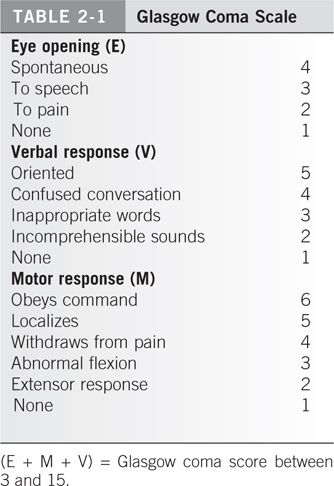
4. Disability. A comprehensive neurologic evaluation should be performed, including evaluation of level of consciousness using the Glasgow coma score, cranial and peripheral nerve function and motor and sensory function. This should be repeated in the primary and secondary survey. Any deterioration in serial exams should prompt neurology or neurosurgical evaluation.
5. Exposure. The patient should be fully undressed to perform a thorough evaluation. They should subsequently be covered in warm blankets, and body temperature should be maintained with warm room temperature, warming blankets or pads, and by infusing warmed IV fluids.
B. Trauma Imaging. Recall that the trauma series was being taken in the trauma room while the primary survey was being conducted. Now that the primary survey has been performed and the most critical steps have been taken, even before a thorough history and physical examination, this trauma imaging series should be reviewed; the examiner is ruling in or out the next most critical clues to saving life and limb. The trauma series classically consisted of three X-rays: lateral cervical spine, an anteroposterior chest, and an anteroposterior pelvic view. However, spine and trauma surgeons have found greater utility in a diagnostic CT scan of the cervical spine as it affords better sensitivity and specificity than radiographs alone and eliminates the need for repeat imaging as often as necessary in the multitrauma patient.9 Any patient who is involved in high-energy trauma, who has head injuries, is under chemical substance influence, or is otherwise deemed unable to provide reliable responses during the primary survey should have this imaging because physical examination can be unreliable.
In circumstances when the cervical spine radiograph is performed instead of the CT scan, such as in alert patients with isolated injury, the images must show the inferior endplate of cervical vertebrae 7 (C7), or it should be deemed inadequate and repeated. Both odontoid and C7–T1 pathology are frequently missed injuries even after the secondary survey. If a spine fracture is detected, then a complete spinal series including anteroposterior, lateral and odontoid cervical views, and thoracic plus lumbar spine view is mandatory in view of the increased possibility of segmental spinal injury. CT may be required to rule out upper cervical fractures. The documented incidence of multiple level spine fractures is 7% to 12%. A full spine series should be obtained in the unconscious trauma victim.
All the X-rays should be taken with excellent technique so as not to obscure bone or soft tissue detail. Care must be taken not to be misled by overlying backboards, over- and under-penetrated films, and equipment, clips, and buckles that are frequently left on the X-ray field. Examples abound of subtle femoral neck fractures that were obscured on the X-ray by a belt buckle, a pneumothorax in the upper lobe that was cut out of view due to positioning, or a critical sacral fracture masked by the opacity of a backboard. When these factors are present, radiographs should be repeated.
C. History and physical examination. The history should include a careful account of the accident, a description of the mechanism of injury, and a statement of the degree of violence involved. Concomitant medical disease, drug abuse, and alcoholism should be considered as contributing factors. The transporting paramedic team or member of the accompanying family should be interviewed for these details if the patient cannot reliably give an appropriate history. A useful mnemonic to guide the initial history is the word AMPLE:
A: Allergies
The physician working up an orthopaedic patient should be particularly aware that open fractures should be treated with certain antibiotics to cover the spectrum of bacteria that are at risk for certain types of wounds (see open fractures below). Furthermore, every patient having an orthopaedic operation should receive perioperative antibiotics, making the question of allergies quite germane. A penicillin allergy is the most common.
M: Medications
Medications can influence surgical decision making. They will also tip off the practitioner to important comorbidities and perhaps imply the need for a general medicine consultation prior to surgery. Patients on anticoagulants should have bleeding and clotting parameters checked as it may be prudent to stop such medicines or reverse a coagulopathy prior to surgery.
P: Past illness
Diabetes can influence outcomes of orthopaedic surgery, and heart disease can increase surgical risk. Steroids and nicotine (the use of tobacco products) increase orthopaedic surgical complications as well as outcomes as measured by healing time and healing rates. These risks should be discussed with patients and family members for proper prognostication.
L: Last meal
This is important when considering whether the patient needs to go to the operating room urgently, as the risk of aspiration of food or vomitus is higher postprandial. Most anesthesiologists opt to hold on the administration of anesthesia within 6 to 8 hours of food intake. This concern should not, however, override the emergent nature of certain life- or limb-threatening conditions, which will be discussed below.
E: Events of Injury
Injury circumstances such as height of fall, direction of impact, presence or absence of restraints (seat belt/air bags), extrication time from vehicle, hours in the field, outside temperature, being trapped under heavy objects, smoke inhalation, and many other possibilities are warning flags to the experienced practitioner, which clue in certain medical or orthopaedic conditions and injury patterns.
D. Secondary survey. The secondary survey is a complete physical examination from head to toe. By this juncture, the potentially life-threatening pathology of the ABCs has been addressed, and necessary resuscitation is underway. The patient should be completely undressed for the secondary survey for a most thorough examination.
- Neurologic mental status. The level of consciousness of the patient should first be noted. A brief “disability exam” in an awake patient is a rapid, organized neurologic examination, which documents mental orientation, verbal response to questioning, and response to stimuli. Furthermore, each extremity should be examined for motor and sensory function as well; accurate documentation is crucial because neurologic examinations can reveal progressive deficits. It is imperative that all four extremities be examined and documented. It is good to develop a pattern of examination and stick with that pattern each time for consistency.
In an unconscious patient, a Glasgow coma score is rapidly conducted on the basis of pupil response to light, motor activity, and withdrawal from painful stimuli (Table 2-1). This information is initially obtained by the medics who perform the initial in-the-field evaluation. The Glasgow score is therefore used as the measure of neurologic progress or deterioration. The medics generally also note the position of the patient at the scene of the accident, especially the head, and whether all limbs were actively moving. It is frustrating to the orthopaedic surgeon or neurosurgeon to be asked to evaluate a patient who has been sedated and chemically paralyzed (for intubation/airway control) in the trauma room, particularly when the initial neurologic examination was not properly documented. In general, the use of maximal monitoring and minimal medication is a useful trauma room principle that avoids such frustration by the examiner who relies on accurate neurologic examinations.
2. Head and neck. Carefully palpate skull and facial bones and look for lacerations hidden in the hair. Cranial trauma should raise an immediate suspicion for cervical spine injury given the sudden and violent force it takes to injure the face and cranium. Radiographs of facial bones are difficult to interpret unless previous clinical examination suggests the presence of trauma. The association between cervical spine and head injuries must be emphasized. In a guided fashion with cervical immobility, remove or loosen the C-collar to palpate the posterior cervical spine looking for tenderness or spasm. In a conscious patient, any neck pain or spasm is a cervical spine injury until proven otherwise. In an unconscious patient, the neck must be protected with a hard C-collar until bony injury is ruled out by cervical imaging and physical examination. A benign physical exam by itself is unreliable if there are distracting injuries or if the patient is intoxicated. If a cervical spine injury is diagnosed, appropriate spine consultation should be obtained immediately, and the extremity neurologic examination should be reported and documented.
3. Thorax and abdomen. Although the thorax and abdomen are largely the domain of the general surgeon, the examiner must inspect, palpate, and auscultate the abdomen and thorax to determine possible underlying injury. Hemothorax and pneumothorax often cause preventable death. Therefore, the chest should be examined carefully and the examination repeated frequently. Furthermore, this assessment helps the orthopaedist place musculoskeletal injuries in the broader context of the patient. Abdominal injury is also a common cause of preventable death. The imprint of clothes or a contusion of the abdominal wall from the seat belt suggests an intra-abdominal injury. Airbags have altered patterns of injury in frontal collision.10 Appropriate diagnostic studies should follow the suspicion of injury, and in many centers the spiral “whole body” CT scan of the chest, abdomen, and pelvis has supplanted selective CT scans, ultrasounds, and peritoneal lavage.
4. Pelvis. Low back pain, pubic tenderness, or pain with compression of the iliac crests can indicate a pelvic ring injury. Sequential anterior to posterior compression over the iliac wings can help to discriminate gross pelvic motion. Pelvic fractures may cause severe internal bleeding, and as stated earlier, a patient can easily lose four units of blood after a displaced pelvic fracture.
A rectal examination must be done in all patients with a spine or pelvic injury, both check for bleeding and loss of sphincter tone indicative of neurologic injury. Furthermore, a high-riding prostate also indicates major urologic disruption common to high-energy pelvic fractures in men. An inspection of the urethral meatus for hemorrhage should also be performed, and such a finding is further indication of a genitourinary system disruption. Bloody urine or the inability to void raises the suspicion of a urethral injury, so a retrograde urethrogram should be considered before a catheter is inserted.11 In male patients, blood at the penile meatus or a “high-riding” prostate seen on rectal examination is a clear indication for obtaining a retrograde urethrogram before bladder catheterization. If the catheter does not pass easily, it should not be forced and the urologist should be consulted. If a bladder injury is suspected, then it is essential to insert an indwelling catheter unless the patient is voiding clear urine.
A bimanual pelvic examination is appropriate in female patients to rule out open fractures that can penetrate the vaginal vault. Perineal inspection for integument lacerations should be conducted and in the setting of displaced pelvic fractures should be assumed to represent an open pelvic fracture.
5. Back and spine. Carefully log roll the patient and palpate the entire spine to detect tenderness or defects of the interspinous ligaments. It is very important that a log roll be conducted properly with three assistants controlling simultaneous rotation of the entire body. A fourth assistant should be controlling the cervical spine (while in a hard collar) with gentle traction. An increase in the interspinous distance accompanied by local swelling and/or tenderness may signify injury. Occasionally, ecchymosis or kyphosis can be recognized, and their presence or absence should be documented.
6. Upper and lower extremity examination. When gross deformity and crepitation are present, further examination of the fracture site is not necessary. Otherwise, all four limbs should be palpated thoroughly and each joint placed through a passive range of motion. Look specifically for point tenderness. Any obvious fractures or deformities are splinted, and any open wounds are covered with sterile saline moistened dressings. Dressings over open wounds, particularly over fractures, should not be taken down multiple times by multiple examiners. Such repeated exposures will only increase the rate of infection with each exposure to the contaminated environment.12 A more detailed description of fracture wound management is given later in this chapter. Every diagnosed fracture should have properly centered X-rays of the joint above and below. Circulation of the limb distal to any fracture should be carefully evaluated and documented. A description and presence of all wounds after applying a sterile dressing should be recorded.
III. ORTHOPAEDIC EMERGENCIES AND URGENCIES. Surgical stabilization of fractures is generally not classified as emergent or urgent and typically can be done on a semielective basis. For example, an isolated, closed fracture that is not threatening local blood supply may wait days to weeks. There are many considerations, however, which go into the optimal timing of surgery, and immediate consultation with an orthopaedist clarifies the issue of timing of surgery.
All the emergent entities, and most of the urgent injuries, ultimately have a common denominator: blood supply, or lack thereof. The lack of circulation affects adequacy of tissue oxygenation, and consequently limb or life is threatened. This may occur on a macroscopic level, such as with a hemorrhaging pelvis in which a person’s life is threatened, or on a microscopic basis, such as when end-organ perfusion is cut off, beginning with occlusion of the venules in a muscle bed due to increased interstitial pressure exceeding intravenous pressure during the condition of compartment syndrome. Threatened blood supply to local tissues can be a more subtle phenomenon that requires further understanding of the vasculature to certain bones. For example, a relatively benign appearing X-ray of a femoral neck fracture to the inexperienced eye may not gain much attention, but the experienced clinician knows that even a nondisplaced femoral neck fracture can threaten the hip joint forever through a process called avascular necrosis (AVN). Certain other orthopaedic injuries may not accurately be classified as emergent because life or limb is not immediately at risk, but they still warrant heightened attention. Such injuries may be classified as urgent because they need prompt action by an orthopaedist and surgical timing in the range of 6 to 24 hours. In the next two sections on emergent and urgent orthopaedic injuries, the discussion will address these in descending order from most to least acute.
A. Orthopaedic emergencies
- Hemodynamically unstable patient with a pelvic fracture. This is the one injury in which circulation can be compromised to the extent that a life is immediately at risk and in which an orthopaedic intervention can save such a life. The pelvic ring can be disrupted in high-energy accidents (or low-energy falls in osteoporotic patients) and nearly always is disrupted in at least two points around the ring. The saying, “it is impossible to break a ring at a single point” nearly always applies to the pelvis. Therefore, the examiner should look for a lesion posteriorly in the sacrum or sacroiliac joint and anteriorly in the pelvic rami or pubic symphisis.
When a pelvic fracture is recognized on the anteroposterior X-ray view obtained with the initial trauma series, two more radiographs should be obtained: a pelvic inlet and pelvic outlet view. These are orthogonal views of the pelvis, which help to critically evaluate all the pelvic bony landmarks as well as displacement of fractures. If there is significant displacement (more than 5 mm) at any one pelvic fracture line, a pelvic CT scan should be obtained. Many orthopaedists will prefer a CT scan with even lesser displacements to more critically evaluate the injury or preoperatively plan. If a fracture line enters the acetabulum, then Judet X-ray views should be obtained. These are 45° angled X-ray views from the right and left sides of the patient centered on the pelvis, once again giving the examiner orthogonal views to critically assess the bony landmarks of each acetabulum. Note that it is wasteful to obtain “five views of the pelvis” for every pelvic fracture as the Judet views are not needed unless the acetabulum is involved. Likewise, inlet and outlet X-rays are not needed unless the pelvic ring is disrupted.
The pelvis is like a cylinder or sphere of bone that contains many critical soft tissue structures and organs such as the bladder, the iliac vessels, prostate or vaginal vault, and the rectum. All these organs are at risk, but the worrisome life-threatening hemorrhage is what must be diagnosed promptly and addressed. Bleeding typically continues until tamponade can occur and clotting factors take control. A sheet or commercial binder around the pelvis of a patient, who is hemodynamically unstable until the anteroposterior radiograph of the pelvis rules in or out a displaced pelvic fracture, is an important measure. The sheet must be clamped very snug at the level of the greater trochanters in order to close down the volume of the broken and separated sphere, thus leading to earlier tamponade of bleeding vessels.13 There is nothing to lose because if the patient does not have such an injury, the binder is simply removed. Some pelvic slings now have pressure calibration to ensure adequate yet safe pressure application through the binder. There is essentially no role for the trauma room application of an external fixator because this maneuver has been obviated by the pelvic sling concept.
2. Extremity arterial injury. Probably, the next most emergent condition that an orthopaedist faces is the extremity that is at risk for limb loss. This can occur due to a torn or lacerated artery or compartment syndrome. Arterial injury can be caused by blunt or penetrating trauma. There are four “hard signs” of arterial injury that warrant immediate vascular exploration, and time should not be wasted ordering and performing a diagnostic arteriogram.14 The rationale is that a vascular surgeon knows the proximity of the injury based on the wound or the X-ray that demonstrates the pathology. There is no sense in using precious minutes finding out what is already known when irreversible ischemic damage to nerve and muscle tissue occurs after 4 hours of warm ischemia time. A warm ischemia in excess of 6 hours is the generally accepted time interval within which arterial continuity must be restored in order to avoid loss of limb.15
The Four “Hard Signs” of Arterial Injury:
a. Pulsatile hemorrhage
b. Expanding hematoma
c. Audible bruit
d. Pulseless limb
The only time an arteriogram would be warranted in such an acute circumstance is when there is multilevel injury (multiple fractures or shotgun wound) in which the vascular surgeon cannot be sure at what level the arterial damage has occurred.
The more difficult diagnostic problem occurs in the majority of patients who present with more subtle clues to vascular injury. Such “soft signs” might include a history of severe hemorrhage at the accident scene, subjectively decreased pulses, a deficit of an anatomically related nerve, or a nonpulsatile hematoma. Other soft signs include the orthopaedic injury patterns that have been associated with a high incidence of arterial damage:
a. Knee dislocations
b. Highly displaced tibia plateau fractures
c. Medial tibia plateau fractures
d.
Related posts:


Stay updated, free articles. Join our Telegram channel

Full access? Get Clinical Tree


