and François Moutet2
(1)
Marseille, France
(2)
Grenoble, France
2.1 Bone Structure
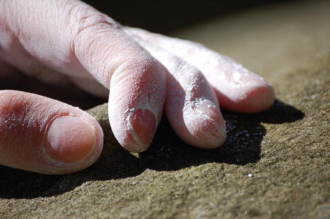
Photo 2.1
2.1.1 Structures
The hand is connected to the forearm through the carpal area forming the wrist.
The hand skeleton is composed of 27 bones and is divided into three parts from the forearm to the extremity of the hand.
The carpals which form the skeleton of the wrist
The metacarpals which form the palm
The phalanges which form the fingers
The hand has two sides: the palm and the dorsum.
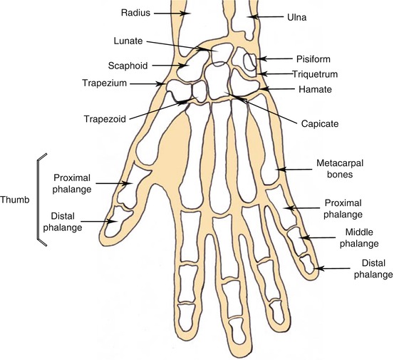
Schema 2.1
Hand bones: palmar side, right hand
2.1.1.1 The Carpals
The carpals include eight bones divided into two areas:
The proximal area which is composed of the scaphoid, the semilunar bone, the triquetrum, and the pisiform bones
The distal area which is composed of the trapeze, the trapezoid, the capitate bone, and the hamate bone
Due to their constitution – a transversal bending with an ulnar-radial direction, concave on the palm, and convex on the back of the hand, covered by flexors and extensors – the carpals are quite mobile.
2.1.1.2 The Metacarpals
The palm is structured by five long bones which are numbered from one to five starting from the thumb metacarpals or the first finger, which is also the most mobile of these five bones.
Each metacarpus is divided into three parts:
The base in the proximal area
The body
The head in the distal area
The metacarpal base is slightly cuboid in shape. The proximal face is connected to one or several bones from the carpal distal area.
The metacarpal head is rather circular and is made of spongy bones covered with cartilage.
The metacarpal body is like a triangular prism made of compact bone tissue.
2.1.1.3 The Phalanges
Each finger is divided into three phalanges except for the thumb with only two phalanges. The hand is consequently composed of 14 phalanges including:
Five proximal phalanges (P1)
Four middle phalanges (P2) for the long fingers
Five distal phalanges (P3/P2) for the thumb
Their size gradually decreases as you get closer to the distal area, and just like the metacarpals, their structure includes a base, a body, and a head.
The first phalange (P1) is called the proximal phalange, the second one (P2) middle phalange, and the third one (P3) distal phalange.
These long bones also have a convex back side and a concave palm side. The third phalange ends up in the phalangeal tuft.
2.1.1.4 The Thumb
The thumb has to be regarded as a proper finger whose main function is to create an opposition in order to act just like digital pliers.
The first phalange can move according to two axes: getting forward or backward the palm or the fingers. Contrary to the first phalange, the second phalange, just like the other fingers, is limited to one movement.
2.1.2 Finger Mechanics
When something is seized, the fingers are the first to move – clinging to the object – before being finally followed by the thumb.
The movement is reversed when something is released – the thumb relaxes first.
In a rest position, the fingers are folded in an inward arch. The little finger which is more convex than the index is also directed toward the thumb.
When fists are clenched, the first finger to fold back is the little finger followed by the ring finger and so on. The movement is reversed when the hand is opened. In fact, each unfolded finger tends to set up a chain reaction.
2.2 Joint Structures
A joint is a sliding area between two bones. Smooth surfaces such as the joint cartilage and the oily liquid – synovia – make the sliding easier. As regards the two bones, they are connected to each other by ligaments.
The finger is joined by three mobile segments. Its position results from a complex balance between several systems:
The passive control – joints and capsular ligaments
The semi-passive and multiple active control – superficial common flexors (FDS) and deep common flexors (FDP)
Therefore, any lesion in that complex system may lead to a dysfunction in the finger.
As regards finger folding, three joints are involved:
The metacarpophalangeal joint (MCP)
The proximal interphalangeal joint (PIP)
The distal interphalangeal joint (DIP)
Their actions differ according to the need. In that precise case, MCP is used up to 77 %, PIP 20 %, and DIP 3 %.
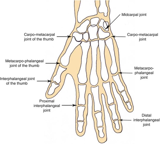
Schema 2.2
Joints: palmar side, right hand
2.2.1 Mediocarpal Joints
These joints connect the three main bones of the proximal area (scaphoid, Lunate, and triquetrum bones) to the four bones of the distal area (trapeze, trapezoid, capitate, and hamate).
A thin and loose capsule is mainly present in the dorsal side, whereas it is more powerful in the front.
Palmar, dorsal, and lateral ligaments keep the whole structure stable.
2.2.2 Carpometacarpal Joints
The long finger’s carpometacarpal joints are barely mobile and are maintained by the palmar and dorsal hard ligaments.
2.2.3 Intermetacarpal Joints
These stiff joints are fixed by the dorsal, palmar, and interosseous metacarpal ligaments.
2.2.4 Thumb Carpometacarpal Joints (Metacarpal and Trapezoid)
It’s a saddle joint allowing the thumb to abduct (to go vertically), to adduct (to go horizontally), or to resist. The saddle joint accounts for the fact that two areas (concave and convex) fit together.
2.2.5 Finger Joints
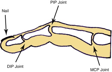
Schema 2.3
Finger joints
2.2.5.1 Metacarpal Phalangeal Joints (MCP)
The metacarpals’ heads and the first phalanges’ bases are the articulated elements.
The lateral ligaments reduce mobility. These joints are surrounded by loose capsules reinforced by palmar ligaments and fiber cartilages.
The MCP joint (condyloid type) is able to move laterally or vertically. The four fingers can follow two axes: flexion extension or abduction (the fingers are wide apart) and adduction (the fingers are close together). Abduction movements are practically impossible with flexed fingers.
2.2.5.2 Proximal Interphalangeal Joints (PIP)
This joint belongs to the hinge joint type – pulley joints – and can only execute a flexion/extension movement on a sagittal plane.
The PIP roughly looks like the knee joint. Lateral ligaments, radial collateral ligament (RCL), and ulnar collateral ligament (UCL) stabilize the joint. The joint capsule is thin at the PIP level and hardly interferes in the stability.
The PIP can only perform one flexion/extension movement whose average angle is 0–100°. It is also more fragile and sometimes quite tricky leading to traumas more serious than in the MCP.
Both the second and third phalanx can fold back onto the next one on a maximum angle of 90°, whereas the first phalange’s degree of mobility is higher (+45° up to +90°) especially when the other phalanges are folded.
2.2.5.3 Distal Interphalangeal Joints (DIP)
This joint connects the second phalanx (P2) to the third one (P3) and presents the same constitution of the PIP.
2.3 Muscle and Tendon Structures
2.3.1 What Is a Tendon?
It’s usually a white circular fiber ending. At each contraction, this resilient non-stretchable fiber, used to fix the muscle on the bone, allows the latter to move. Tendons are extremely long since the muscles at the origin of each movement are situated at half distance of the proximal area of the forearm.
2.3.2 Various Muscles of the Hand
2.3.2.1 Finger Flexors
Flexor Digitorum Superficialis (FDS)
This muscle comes from two points:
The humeral fascicle of muscle fibers (the main source) which ends up in the epitrochlea, that is to say, the lower part of the humerus, and in the ulnar coronoid process of the ulnaRelated posts:

Stay updated, free articles. Join our Telegram channel

Full access? Get Clinical Tree


