Tarsometatarsal Lisfranc Injuries: Evaluation and Management
Bruce J. Sangeorzan
Stephen K. Benirschke
Mark T. Gould
Indications/Contraindications
The primary surgical indication for treatment of an injury is the knowledge that the injury will do poorly with nonoperative treatment. Conceptually, tarsometatarsal injuries that will lead to a loss of the arch or significant deformity if treated conservatively should be treated surgically; these include both displaced injuries and subtle injuries that have instability in two planes. The decision to treat a tarsometatarsal injury surgically is based on both physical examination and radiographic studies.
The transverse and longitudinal arches of the foot depend on the tarsometatarsal joints to make the foot sufficiently rigid to support the body, much as the apical blocks of ice support an igloo. Unstable tarsometatarsal injuries that compromise this structural integrity may result in deformity of the foot. In the majority of displaced injuries, the metatarsals displace dorsally and laterally on the tarsal bones, which produces pes planus with forefoot abduction. As a result, when weight is borne on the foot, it collapses. During heel lift, further deforming forces that act on the midfoot tend to exacerbate the deformity. For the metatarsals to displace in this direction, the plantar tarsometatarsal (Lisfranc) ligaments must be disrupted. Operative treatment is indicated when an ambulatory patient has an injury that renders the foot mechanically unsound, deformed, or both.
Ambulatory patients with displaced Lisfranc joints that are apparent on the plain x-rays and who have all the stabilizing ligaments disrupted are candidates for surgery. When the injuries are subtle or apparently nondisplaced, operative treatment is indicated only when two-plane instability is detected on clinical examination or stress x-rays. Because the foot functions in weight bearing, the integrity of the plantar ligaments is of greater importance than that of the dorsal ligaments.
Contraindications to surgical intervention include nonambulatory individuals, patients with serious vascular disease unlikely to heal a surgical incision but who have no significant deformity, or an injury that is unstable in only the transverse plane. Lisfranc injuries with only bone injuries can be treated by closed means or by closed reduction with percutaneous pinning. When deformity and compromised circulation is found, the surgeon faces a dilemma. Leaving a deformity puts the patient at risk for ulceration, while treating it surgically puts the patient at risk for wound-healing problems. In this circumstance, a vascular surgeon may be consulted to evaluate whether an inflow procedure would be beneficial prior to orthopedic intervention.
Neurologic impairment is also a cause for concern. The physician must decide whether sufficient energy produced the injury or whether an underlying neuropathic condition exists. Trivial injuries that cause significant displacement should stimulate an investigation into a possible neuropathic condition. A Charcot neuropathic foot has different indications for treatment and calls for different technique than does a foot without preexisting neuropathy. For treatment of a Lisfranc injury in the presence of peripheral neuropathy, more fixation will be required, and a longer period of postoperative protection is indicated.
Physical Examination Criteria
Lisfranc injury is often missed and is one of the few injuries in orthopedics in which the maxim “the eye doesn’t see what the mind doesn’t search for” is most appropriate. Swelling and tenderness in the midfoot with no obvious fracture should trigger a high index of suspicion. Instability should be determined by physical examination. The physician grasps the metatarsal heads and applies a dorsal force to the forefoot while the other hand palpates the tarsometatarsal joint. Dorsal subluxation or dislocation of the bases of the metatarsals suggests instability (Fig 36.1). If the first and second metatarsal can be displaced medially or laterally as well, global instability is present, and surgical treatment is needed. Low-energy injuries interrupt the medial capsule but do not disrupt the plantar ligaments. When the
plantar ligaments are intact, no dorsal subluxation will occur with stress examination. These injuries may be treated nonoperatively or with less rigid fixation at the discretion of the examining surgeon.
plantar ligaments are intact, no dorsal subluxation will occur with stress examination. These injuries may be treated nonoperatively or with less rigid fixation at the discretion of the examining surgeon.
 Figure 36.1. A–C. Diagrammatic representation of a dorsal view of the foot. The metatarsal bases are forced laterally and dorsally. |
Radiographic Criteria
With ligamentous disruption of the midfoot without fracture, x-rays made while the patient is not weight bearing may be deceptively benign. The ligaments are torn with initial displacement; however, when the deforming force is removed, the foot may spring back into a neutral position, concealing gross instability. The physician should be suspicious whenever midfoot gross swelling and pain is found.
When disruptions are found on several radiographic plane films, subtle tarsometatarsal injury is suggested. The first and most reliable image shows disruption in the continuity of a line drawn from the medial base of the second metatarsal to the medial side of intermediate cuneiform on the anteroposterior (AP) and oblique views (Fig. 36.2A).
The second most reliable type of image shows widening of the interval between the first and second ray; these x-rays should arouse suspicion. If tenderness is evident upon palpation, stress views should be obtained.
On a third most important observation, the medial side of the base of the fourth metatarsal should line up with the medial side of the cuboid on the oblique view. This is a soft sign because the cross section of the metatarsal base is not equal to the cross section of the cuboid. As a result, a step-off may be present if the angle of the beam is slightly misdirected. On the lateral view, the metatarsals are aligned with the cuneiforms at the dorsal cortex. When ligament injury is extant, the metatarsals are typically dorsally displaced in relation to the cuneiforms.
Finally, any disruption of the medial column line (MCL), a line tangential to the medial aspect of the navicular and medial cuneiform, can be found on two views. The disruption will show on the intersection of the base of the first metatarsal on an AP view taken during weight bearing. In addition, views taken during abduction stress reliably predict disruption of the Lisfranc ligamentous complex.
Preoperative Planning
Physical examination should document the status of the dorsalis pedis and posterior tibial pulses, the integrity of the skin, and the habitus of the foot. Tendon entrapment may be demonstrated by an altered, uncorrectable position of the toes or midfoot. Intact or altered sensation should be documented.
Preoperative imaging should include a simulated AP and lateral view of the foot under weight bearing, as well as an oblique view. Oblique views are essential in evaluating a midfoot injury and should be included in the foot trauma series. If the presence, location, or degree of injury is uncertain, stress x-rays should be taken in two planes. Typically they are done through use of fluoroscopy so the surgeon can make certain that the correct plane is achieved for the image. When the index of suspicion is high, the stress roentgenogram is performed in an operating room (OR), so that if the injury is confirmed, surgery can be done under the same anesthetic.
An appropriate anesthetic is given, and the fluoroscopy unit is brought into the OR suite. The table is bent at the knees so the foot is relatively parallel to the floor. While wearing lead gloves, the surgeon grasps the first and second metatarsal heads with one hand and the hind foot with the other. With the thumb placed over the cuboid to act as a fulcrum, the forefoot is abducted and an AP x-ray is obtained. Instability is present if a gap occurs on the medial side of the first or second tarsometatarsal joint, or disruption of the MCL is produced (see Fig. 36.2D).
Stress views in the lateral plane are performed if uncertainty exists. This is done with the surgeon grasping the midfoot with one hand and the forefoot with the other, and acutely plantarflexing through the tarsometatarsal joint. The fluoroscopy unit is oriented across the
table and an image is obtained. Although the tarsometatarsal joints may angulate, they should not open asymmetrically. Subluxation indicates that the joints are unstable.
table and an image is obtained. Although the tarsometatarsal joints may angulate, they should not open asymmetrically. Subluxation indicates that the joints are unstable.
 Figure 36.2. A. An AP x-ray demonstrating a subtle Lisfranc injury. The base of the second metatarsal is displaced laterally. B. This lateral x-ray, taken under nonweight-bearing conditions, shows that the dorsal cortex of the second metatarsal is subluxed dorsally relative to its cuneiform. C. A scout view is used to confirm that the foot is in the correct position for assessing the tarsometatarsal joints. D. The stress x-ray reveals instability in the first, second, and probably third tarsometatarsal joints. E. An intraoperative fluoroscopic image taken after fixation reveals that the third metatarsal is stable. F. Six weeks following surgery, the reduction appears anatomic and the clinical position of the foot is good. G. Alignment of the metatarsal bases is restored in both planes. |
Possible instability should be investigated at the intercuneiform level (Fig. 36.3). These injuries are not rare, but because they are subtle, out of the plane of standard x-rays, and not well described, they are easily missed. Treatment follows the same principles as those at the Lisfranc level. Stress views in the AP plane are used to confirm the injury. Because little motion is characteristic of most of the intercuneiform joints, any significant motion is abnormal. If the instability is great enough to allow subluxation of the midfoot, it should be treated. Displacement of the intercuneiform joints leads to deformity that is poorly understood and difficult to treat.
If x-rays are done in AP, lateral, and oblique planes, and stress views are obtained when there is uncertainty, additional imaging modalities should not be necessary. Computed tomographic (CT) scans of the midfoot are difficult to interpret. The role of the magnetic resonance imaging (MRI) scan has not been established.
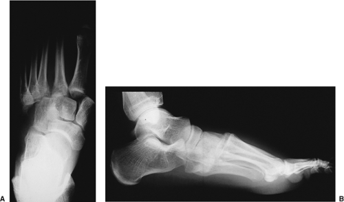 Figure 36.3. A. An AP x-ray of a left foot with severe Lisfranc injury. All five metatarsals are displaced laterally. B. The lateral view, taken under nonweight-bearing conditions, shows a dorsal dislocation. |
Surgery
Timing
Related posts:
 Clavicular Fractures: Open Reduction Internal Fixation
Olecranon Fractures: Open Reduction Internal Fixation
Femoral Neck Fractures: Arthroplasty
Tibial Pilon Fractures: Open Reduction Internal Fixation
Posterior Pelvic-Ring Disruptions: Iliosacral Screws
The Treatment of Distal Tibia Peri-articular Fractures with Circular Ring Fixators
Clavicular Fractures: Open Reduction Internal Fixation
Olecranon Fractures: Open Reduction Internal Fixation
Femoral Neck Fractures: Arthroplasty
Tibial Pilon Fractures: Open Reduction Internal Fixation
Posterior Pelvic-Ring Disruptions: Iliosacral Screws
The Treatment of Distal Tibia Peri-articular Fractures with Circular Ring Fixators
Stay updated, free articles. Join our Telegram channel

Full access? Get Clinical Tree


