David P. Barei
Sean E. Nork
Sterile Instruments/Equipment
- Tourniquet if desired
- Headlight
- Small pointed bone reduction clamps (Weber clamps)
- Dental picks and Freer elevators
- Schanz pins (2.5 to 4.0 mm)
- Femoral distractor (small)
- Micro-oscillating saw if medial malleolar osteotomy needed
- 3.5- to 4.0-mm cancellous and cortical screws for osteotomy fixation
- Implants
- Mini- and small-fragment screws and mini-fragment plates and screws (2.0/2.4/2.7/3.5 mm)
- Autograft, cancellous allograft or other bone substitute for structural defects
- Mini- and small-fragment screws and mini-fragment plates and screws (2.0/2.4/2.7/3.5 mm)
- K-wires and wire driver/drill
Positioning and Imaging
- Supine at the foot end of the radiolucent cantilever table.
- Small bump underneath the ipsilateral hip.
- Three views are used to assess talar fracture reductions: lateral of talus/ankle, Canale view, and ankle mortise.
Surgical Approaches
- Simultaneous medial and lateral approaches.1
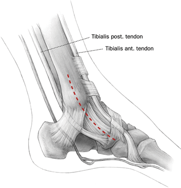
Figure 23-1. Plane of an anteromedial incision. (From Vallier HA, Nork SE, Benirschke SK, et al. Surgical treatment of talar body fractures. J Bone Joint Surg Am. 2004;86: 180–192. Reprinted with permission.)
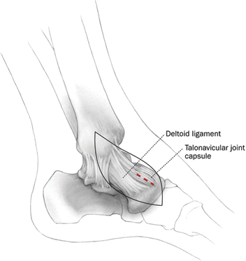
Figure 23-2. Deep dissection for an anteromedial approach. (From Vallier HA, Nork SE, Benirschke SK, et al. Surgical treatment of talar body fractures. J Bone Joint Surg Am. 2004;86:180–192. Reprinted with permission.)
![]()
- From anterior medial malleolus to navicular.
- Deltoid protected.
- Plantar dissection avoided.
- Incise the talonavicular joint capsule, proximal to medial tarsal artery.
- Expose the dorsomedial talar neck and medial body.
- Visualization includes medial talar dome, medial talar neck, and talar head.
- Retraction of capsule and dorsal soft tissues allows visualization of the anterior aspect of the talar dome to the lateral side.
- Plan incision to allow medial malleolar osteotomy depending on the fracture pattern.
- Retraction of capsule and dorsal soft tissues allows visualization of the anterior aspect of the talar dome to the lateral side.
- Anterolateral incision is in line with the fourth ray of foot (Figs. 23-3 and 23-4).
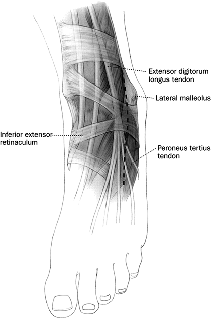
Figure 23-3. Incision for an anterolateral approach for a talus fracture. (From Vallier HA, Nork SE, Benirschke SK, et al . Surgical treatment of talar body fractures. J Bone Joint Surg Am. 2004;86:180–192. Reprinted with permission.
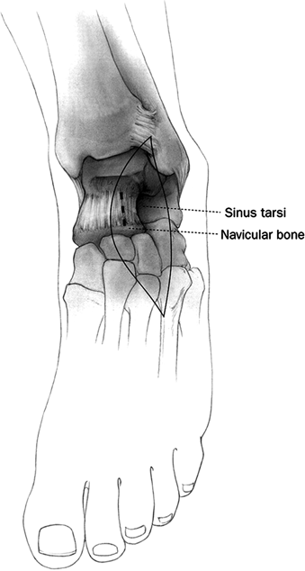
Figure 23-4. Deep dissection for an anterolateral approach to the talus. (From Vallier HA, Nork SE, Benirschke SK, et al. Surgical treatment of talar body fractures. J Bone Joint Surg Am. 2004;86:180–192. Reprinted with permission.)
![]()
- Sharp dissection, full thickness flaps.
- Avoid superficial peroneal nerve in proximal incision.
- Develop interval between long toe extensors and extensor brevis musculature.
- Brevis musculature typically retracted plantar/lateral.
- Avoid branches of dorsalis pedis, muscular branches to EHB and EDB, and lateral tarsal artery.
- Brevis musculature typically retracted plantar/lateral.
- Retract anterior compartment tendons medially.
- Reflect or excise sinus tarsi fat.
- Visualization should include the lateral dome of the talus, lateral process of talus, lateral talar neck, lateral talar head, and the talofibular articulation.
- Dissection plantar to the lateral process of the talus allows access to the posterior facet of the subtalar joint, facilitating debridement of osseous and chondral debris.
Reduction and Fixation Tips
Talar Neck
- Use cortical interdigitations for reduction; more commonly this is on the lateral side or inferomedially, as impaction/comminution is typically dorsomedially.
- Some vertical neck fractures traverse the anterior chondral surface of the talar body.
- Often the chondral interdigitations along this portion of the fracture provide an excellent assessment of reduction accuracy.
- Visually assess the quality of reduction, medially and laterally using both incisions.
- Subtle rotational and angulatory malreductions can be appreciated from one side or the other.
- Radiographic correlation requires understanding of the normal talar relationships with the rest of the foot.
- Talar axis on the AP view.
- Common malreduction deformity is varus.
- Talar axis on the lateral view.
- Common malreduction deformity is extension, especially in high energy fractures.
- These high energy injuries often require tricortical or cancellous bone graft to “strut” the area of impaction and comminution (dorsally) so as to restore the talar-first metatarsal angle.
- The talar-first metatarsal angle should be nearly parallel on the AP and lateral radiographs.
- Common malreduction deformity is extension, especially in high energy fractures.
- The anterior and middle facets are on the talar head fragment; the posterior facet is on the body fragment.
- Check to ensure that these are congruent radiographically.
- Talar axis on the AP view.
- Managing circumferential comminution
- Difficult problem.
- Small external fixator from the distal medial tibia to the medial aspect of the navicular and from the fibula to the lateral aspect of the navicular will allow controlled restoration of talar neck length and angulation.
- Radiographically reduce the anterior and middle facet (i.e., head fragment) and the posterior facet (i.e., body fragment) to the calcaneus.
- If these joints are congruent, then the talar neck length is correct.
- Difficult problem.
Provisional Fixation
- Multiple retrograde K-wires from both the anteromedial and anterolateral exposures.
- Avoid placing wires in the fibula or across the tibiotalar/subtalar articulations.
- A femoral distractor with Schanz pins in the medial distal tibia and the cuneiforms can be used to aid in fracture visualization and reduction (Fig. 23-5).
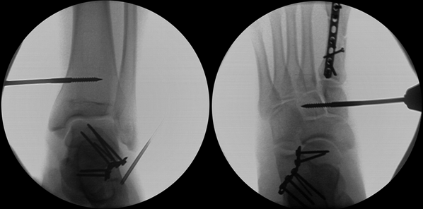
Figure 23-5. A universal distractor placed from the tibia to the midfoot can restore talar neck length.
Stay updated, free articles. Join our Telegram channel

Full access? Get Clinical Tree


