Surgical Fixation of Metacarpal Fractures
Patient Selection

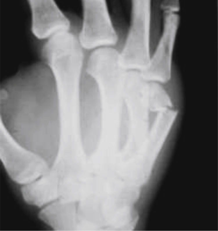
Figure 1Oblique radiograph of the hand demonstrates a metacarpal neck fracture of the small finger (boxer’s fracture). These fractures typically are seen in a patient who has punched a solid object.
Metacarpal fractures most commonly result from an axial load transmitted from metacarpophalangeal (MCP) joint proximally down shaft of metacarpal (Figure 1)
Most metacarpal fractures are treated nonsurgically
Indications—Extensive soft-tissue injury, multiple metacarpal fractures, isolated metacarpal fractures with rotational deformity, angular deformity, or excessive shortening
Contraindications—None
Preoperative Imaging
AP, lateral, and oblique radiographs of the hand
Use Brewerton radiographic view to assess metacarpal head fractures
Procedure
Special Equipment
Mini C-arm fluoroscopy
Kirschner wires (K-wires) plates may be helpful (2.0-2.7 mm size)
Surgical Technique
Metacarpal Neck Fractures

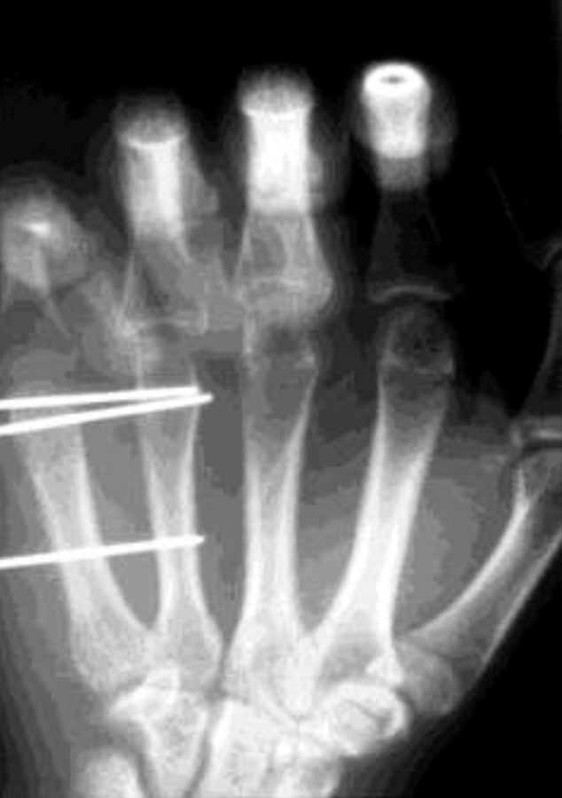
Figure 2Fluoroscopic PA image shows a fifth metacarpal neck fracture that has undergone closed reduction and stabilization with cross-pinning of the proximal and distal fragments to the fourth metacarpal.
Many methods of fixation are recommended
May place K-wires transversely across metacarpal neck into adjacent metacarpal; easier for border digits (Figure 2)
May place K-wires longitudinally down metacarpal
When pinning distal to proximal, flex MCP joint to control distal fragment; use smooth 0.045- or 0.054-in K-wires; start on radial or ulnar collateral recess
Reduce fracture as wire approaches fracture site; advance wire into base; consider advancing with mallet to avoid cortical penetration
Place K-wires with bouquet technique for index or small digit
For index, make 2-cm skin incision on radial side of second metacarpal base
For small digit, make incision on ulnar side
Elevate extensor tendon insertion
Cut off tip of 0.045-in K-wire; bend gently along length
Under fluoroscopy, identify proximal part of metaphysis; penetrate through canal with 2-mm drill; enlarge to 5 mm as needed
Place precontoured K-wire into metacarpal base and advance across fracture; place multiple K-wires similarly to enter distal fragment at various sites and create bouquet
Stay updated, free articles. Join our Telegram channel

Full access? Get Clinical Tree


