Forefoot Supinatus
Erica L. Evans, DPM∗eevans1@wpahs.org and Alan R. Catanzariti, DPM, Foot & Ankle Institute, Foot & Ankle Surgery, West Penn Hospital, 4800 Friendship Avenue, N1, Pittsburgh, PA 15224, USA
The supination of the forefoot that develops with adult acquired flatfoot is defined as forefoot supinatus. This deformity is an acquired soft tissue adaptation in which the forefoot is inverted on the rearfoot. Forefoot supinatus is a reducible deformity. Forefoot supinatus can mimic, and often be mistaken for, a forefoot varus. A forefoot varus differs from forefoot supinatus in that a forefoot varus is a congenital osseous deformity that induces subtalar joint pronation, whereas forefoot supinatus is acquired and develops because of subtalar joint pronation. This article discusses the acquired form of forefoot supinatus.
Key points
The adult acquired flatfoot deformity (AAFD) is one of the most common disorders seen by foot and ankle specialists. A flatfoot is characterized by abduction and supination of the forefoot with a collapsed medial longitudinal arch. However, the relationships between the osseous and soft tissue structures that contribute to a deformity often vary in degree of severity, nature, and location of pain and instability.1
The supination of the forefoot that develops with adult acquired flatfoot is defined as forefoot supinatus. This deformity is an acquired soft tissue adaptation in which the forefoot is inverted on the rearfoot. The degree of forefoot supinatus present in an individual patient is the function of calcaneal eversion, the chronicity of the problem, and adaptive muscle and osseous changes. When significant adaptive alteration in muscle, ligament, and articular surfaces has developed over an extended period of time, the supinatus may become fixed and difficult to differentiate from forefoot varus.2
Etiology
Supination of the foot around the longitudinal axis of the midtarsal joint is required to accommodate normal calcaneal eversion in gait (4–6 degrees). When excessive calcaneal eversion is present, excessive inversion of the foot about the longitudinal midtarsal joint axis is the usual corollary finding. In addition to longitudinal axis supinatory subluxation, dorsal displacement of the first ray also occurs with calcaneal eversion as the result of first ray hypermobility.2
Forefoot supinatus is a compensatory deformity that develops secondarily to pathologic forces that tend to cause dorsiflexion or prevent plantarflexion of the metatarsals. There are two main mechanisms recognized that contribute to the development of a forefoot supinatus deformity: ankle equinus and excessive subtalar joint pronation.3
Equinus refers to any condition, either structural or functional, that restricts ankle joint dorsiflexion to less than 10 degrees when the subtalar joint is in neutral and the midtarsal joint is fully pronated.4 Compensation for an ankle equinus typically results in subtalar and midtarsal joint pronation. The Achilles tendon forces the calcaneus into valgus and limits subtalar joint inversion in the presence of equinus.1 Ankle equinus ultimately results in a direct dorsiflexory force on the metatarsals after the limit of ankle joint dorsiflexion is reached.3
The second mechanism responsible for the development of forefoot supinatus is excessive subtalar joint pronation during forefoot loading. The pronated position of the subtalar joint allows for increased pronation of the midtarsal joint. Any additional eversion of the forefoot about the oblique axis of the midtarsal joint must be compensated by further forefoot inversion by dorsiflexion of the medial column.3
An unlocked midtarsal joint during midstance allows the concentric contraction of the Achilles tendon to plantarflex the hindfoot on the forefoot. This places significant overload on the posterior tibial tendon and spring ligament, in addition to the long and short plantar ligaments and plantar aponeurosis.5 This process results in the radiographic finding of “lateral peritalar subluxation.”6 This peritalar subluxation results in midfoot abduction and forefoot supinatus.1
Whatever the contribution of each deformity, the result in long-standing cases of calcaneal eversion is a fixed or semi-rigid position of inversion of the forefoot relative to the rearfoot.2 Both the underlying etiologic factor and the acquired deformity should be addressed when treating a supinatus deformity.
Clinical evaluation
Before any specific examination for forefoot supinatus, the range of motion of the subtalar, midtarsal, first ray, and first metatarsal phalangeal joints should be evaluated.2
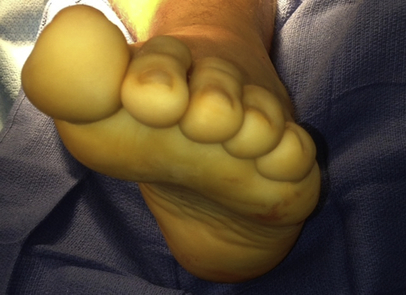
The position of the foot required for measuring the forefoot to rearfoot relationship must be maintained for all subsequent measurements. Deformity through the medial column is best evaluated with the patient in the seated position. The hindfoot is placed in a “neutral position” by centering the navicular over the talar head. The deformity is confirmed by palpating the medial border of the foot and the relationship of the navicular tuberosity to the talar head. As the hindfoot is held in this position with one hand, the opposite hand is used to passively bring the ankle to neutral dorsiflexion by placing force on the plantar aspect of the fourth and fifth metatarsal heads. At this point, the relationship of the first and fifth metatarsals are evaluated by viewing the foot “head-on” to determine the degree of elevation of the first ray relative to the fifth ray (Fig. 1).7

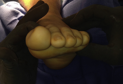
Stability of the first ray is then assessed, again in a seated position, by stabilizing the lesser four metatarsals with one hand and then using the opposite hand to manipulate the first metatarsal. The dorsiflexion and plantarflexion of the first ray in relation to the rest of the foot is assessed while the windlass mechanism is engaged to determine if there is excessive sagittal plane motion or if there is pain or crepitus with range of motion (Fig. 2).8
Stay updated, free articles. Join our Telegram channel

Full access? Get Clinical Tree


