Sterile Instruments/Equipment
- On-table traction or fracture table
- 5.0-mm Schanz pins for manipulative joysticks
- Large pointed bone reduction clamps (Weber clamps)
- Serrated bone reduction clamps
- Femoral distractor
- Ball-spike pushers
- Shoulder hook/bone hook
- K-wires and wire driver/drill
- Reamers
- Implants
- Intramedullary nail
- Reconstruction (cephalomedullary) versus conventional (condylomedullary)
- Proximal femoral locking plate
- Blade plate
- Intramedullary nail
Positioning
- Supine on a radiolucent table.
- Bump under the ipsilateral hemipelvis and torso.
- Move patient as far as possible to the table’s edge so that buttock is partially overhanging the edge of the table, particularly if a piriformis nail is being planned.
- Improves access for nail insertion
- Confirm ability to obtain adequate intraoperative AP and lateral images prior to prepping and draping.
- Improves access for nail insertion
- Skeletal traction through a distal femoral pin is useful in restoring the fracture length.
- Limb elevated on radiolucent foam ramp or triangular wedge.
- Assists in reducing fracture and matching flexion of distal fragment to proximal fragment’s flexion deformity.
- Limb elevated on radiolucent foam ramp or triangular wedge.
Surgical Approaches for Open Treatment
- Standard lateral approach to proximal femur.
- Incise iliotibial band.
- Elevate vastus lateralis origin from vastus ridge; identify perforating vessels.
- Selective approaches for clamp placements or joysticks.
Reduction and Implant Techniques
- The most common deforming forces are flexion, abduction, and external rotation of the proximal fragment due to tendon insertions and muscular forces (Fig. 15-1).
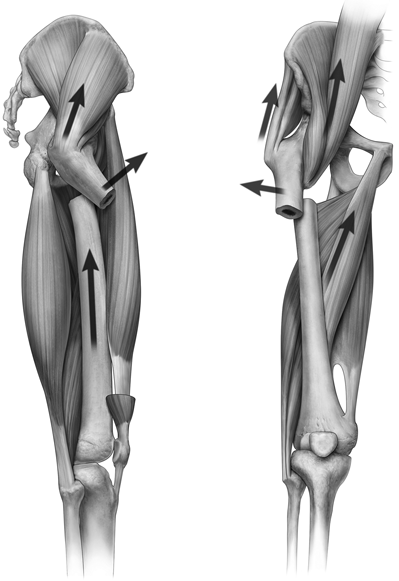
Figure 15-1. The primary forces that must be counteracted with reduction maneuvers in subtrochanteric fractures are flexion, abduction, and external rotation of the proximal fragment.
![]()
- Two main reduction methods can be used separately or in combination.
- Reducing the distal fragment to the proximal fragment without manipulating the proximal fragment.
- This requires matching the flexion, external rotation, and variable abduction with concomitant traction.
- Reducing the proximal fragment to the distal fragment without manipulating the distal fragment.
- This is accomplished by controlling the proximal fragment either directly (open technique) or percutaneously with joysticks, reduction clamps, or other devices.
- In this scenario, the deforming forces on the proximal fragment are counteracted by adducting, extending, and internally rotating the proximal fragment.
- This is difficult to correct with traction alone.
- This is accomplished by controlling the proximal fragment either directly (open technique) or percutaneously with joysticks, reduction clamps, or other devices.
- Reducing the distal fragment to the proximal fragment without manipulating the proximal fragment.
- Limb alignment and fracture reduction must be maintained during reaming and implant placement so as to assure a concentric nail position, while maintaining fracture reduction.
- When placing a trochanteric entry nail with cephomedullary fixation (i.e., reconstruction screws), the starting point should be slightly more anterior and medial than is typical for treating a shaft fracture.
- A properly placed starting point is critical for obtaining and maintaining an acceptable reduction. This starting point is variable and based on the specific implant used. Adequate AP and lateral intraoperative imaging is necessary to confirm perfect entry point placement.
- Occasionally, attempts at translating the reamers medially will displace fracture fragments medially instead of reaming medial bone.
- When reaming with the patient supine, after each reamer is removed, the guide wire will tend to rest in the posterolateral aspect of the reamer track due to guide wire’s trajectory, gravity, and flexion/abduction of the proximal fragment.
- This will cause each successive reamer pass in the proximal fragment to occur more and more laterally and posteriorly if this is not addressed and counteracted (Fig. 15-2).
- Occasionally, attempts at translating the reamers medially will displace fracture fragments medially instead of reaming medial bone.
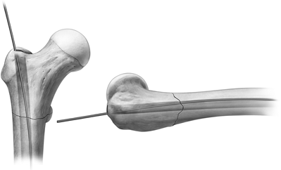
Figure 15-2. Gravity and soft tissue forces will tend to position the guide wire laterally (left) and posteriorly (right). If not corrected, this may cause eccentric reaming of the entry portal with each successive reamer pass.
![]()
Related posts:


Stay updated, free articles. Join our Telegram channel

Full access? Get Clinical Tree


