1
Structure and Function of Cartilage Biology
MICHAEL A. SCHWARTZ AND MICHAEL G. CICCOTTI
Articular cartilage is a highly specialized tissue, exhibiting unique biomechanical properties. It is specifically organized to allow for the complex movements performed by synovial joints. With its specialized structure, articular cartilage has the benefit of excellent load-bearing characteristics, as well as friction, lubrication, and wear characteristics. This design enables articular cartilage to withstand many cycles of stress, a resiliency that is unique to the tissue.
 Composition
The makeup of articular cartilage is ~95% extracellular and 5% cellular, exclusively chondrocytes.1 The vast extra-cellular space is ~75% water, 20% collagen, 5% proteoglycans, and less than 1% other proteins, such as various growth factors, adhesive proteins, enzymes, and lipids.
Extracellular
Interstitial fluid is distributed more densely at the surface of articular cartilage, with a superficial concentration of 85%, and a its deep concentration of 65%.2 This high surface water concentration allows for deformation in response to external stress, by shifting of the fluid in and out of cartilage. In addition, water plays a pivotal role in joint nutrition and lubrication.
The collagen of articular cartilage is primarily type II (90–95%), with small contributions from types IX and XI.3–5 Under electron microscopy, collagen is noted to have a cross-banded fibrillar structure. Three α chains intertwine into a triple helix, forming a procollagen molecule. After the N-and C- terminal ends are cleaved off, the collagen fibrils are fashioned in a quarter-stagger overlapping configuration, producing the cross-banded appearance.6–13 Covalent interfibrillar cross-links form, connecting the collagen molecules. This collagen imparts to the cartilage its mechanical properties of tensile stiffness, strength, and mechanical stability. The individual fiber diameter and orientation, the density of collagen fibers, and the amount of collagen interfibrillar cross-links all contribute to these unique characteristics.4,11–17 In addition, collagen type XI functions as an adhesive that holds the collagen latticework together.18–20
Proteoglycans are glycosaminoglycan aggregates that are produced by the chondrocytes and secreted into the matrix. They are primarily responsible for the compressive strength of cartilage, by regulating the matrix hydration via trapping and holding water. The structure of the proteoglycan aggrecan molecule resembles a test-tube brush with keratin and chondroitin sulfate (glycosaminoglycan) side chains bound to a protein core. Multiple proteoglycan aggrecans are attached to a hyaluronic acid (HA) backbone via a link protein, forming a proteoglycan aggregate (Fig. 1–1). The concentrations of proteoglycans are highest in the articular cartilage middle zones and lowest in the more superficial zone.
Cartilage synthesis is regulated by various protein growth factors. Of these, transforming growth factor-β (TGF-β), fibroblast growth factor (FGF), insulin-like growth factor-I (IGF-I), and platelet-derived growth factor (PDGF) are best understood. It is believed that TGF-p can stimulate proteoglycan synthesis and inhibit type II collagen synthesis. FGF stimulates DNA synthesis in adult articular chondrocytes. IGF-I stimulates DNA and matrix synthesis in both adult articular cartilage and immature growth plate cartilage. PDGF functions to promote proliferative responses on many mesenchymal cell types, including various connective tissues.
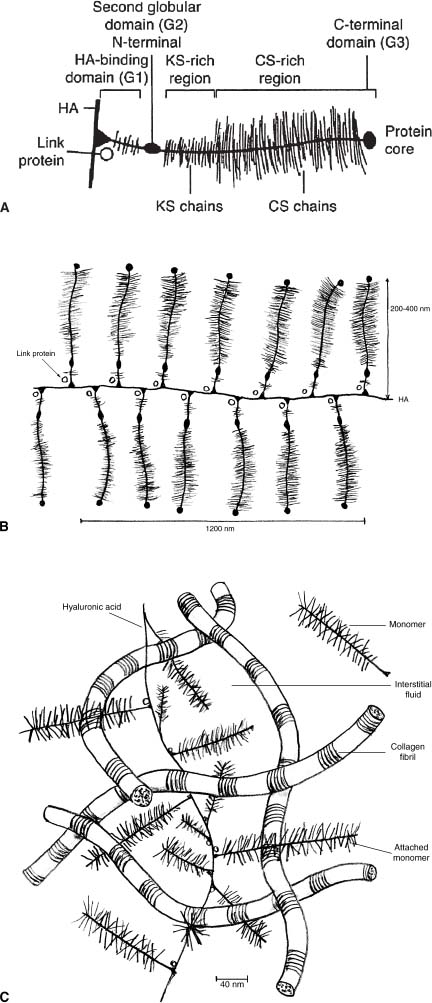
FIGURE 1–1 The structure of aggregating proteoglycans and their association with the collagen matrix. (A) The glycosaminoglycans keratin sulfate (KS) and chondroitin sulfate (CS) attach to aggrecan, which in turn binds to hyaluronic acid (HA) via its N-terminal globular domain through interaction facilitated by link protein. (B) HA is a large polysaccharide composed of repeats of a disaccharide of N-acetylglucosamine and glucuronic acid. (C) The negative charge associated with the proteoglycans trapped in a collagen matrix attracts cations and water into the matrix. (A and B by permission from Mankin HJ, Mow VC, Buckwalter JA, Iannotti JP, Ratcliffe A. Articular cartilage structure, composition, and function. In: Buckwalter JS, Einhorn TA, Simon SR, eds. Orthopaedic Basic Science: Biology and Biomechanics of the Musculoskeletal System, 2nd ed. Rosemont, IL: American Academy of Orthopaedic Surgeons, 2000:449–450. C by permission from Mow VC, Proctor CS, Kelly MA: Biomechanics of articular cartilage. In: Nordin M, Frankel VH, eds. Basic Biomechanics of the Musculoskeletal System, 2nd ed. Philadelphia: Lea and Febiger, 1989:31–57.)
Minimal information is available on the various adhesives and other proteins in articular cartilage. The best understood is link protein, which helps stabilize the proteoglycan aggregates.21,22 Anchorin and chondronectin are examples of other adhesive molecules that are known to exist in articular cartilage. There are also several major and minor cartilage matrix proteins that function to transduce signals from the extracellular environment to the cells via integrins and other similar cell-surface receptors.
Cellular
In mature articular cartilage, chondrocytes occupy only ~5% of the total volume. As a result of this comparatively low cell volume, chondrocytes are considered to be very metabolically active cells (Fig. 1–2).1 They function to regulate the synthesis, maintenance, and turnover of the extracellular matrix proteins.
 Structure
Articular cartilage is arranged in a highly stratified fashion, with four distinct but adjoining layers. Within each layer, there is a specific cellular organization and morphology, as well as varied degrees of tensile strength and stiffness. This ultrastructure is designed to enable each individual layer to fulfill its mechanical role. The four zones are (1) the tangential (superficial) zone, (2) the transitional (intermediate) zone, (3) the radial (deep) zone, and (4) the zone of calcified cartilage (Fig. 1–3).
Tangential (Superficial) Zone
The tangential zone forms the surface of the articular cartilage. It consists mainly of densely packed collagen fibrils, which are secreted by elongated chondrocytes and oriented parallel to the surface. Its high collagen-to-proteoglycan ratio correlates with its particularly high tensile strength and its lack of swelling properties seen in other zones. This imparts to this zone the ability to function against shear.
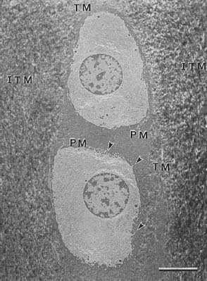
FIGURE 1–2 Electron micrograph of a pair of radial zone chondrocytes embedded in extracellular matrix. The chondrocytes show organelles necessary for matrix synthesis, including endoplasmic reticulum and Golgi’s membranes. The extracellular matrix is compartmentalized into the pericellular matrix (PM), the territorial matrix (TM), and the interterritorial matrix (ITM). Arrowheads: fine cellular processes. Original magnification ×3500; bar = 5 μm. (By permission from Hunziker E, Michel M, Studer D. Ultrastructure of adult human articular cartilage matrix after cryotechnical processing. Microsc Res Tech 1997;37:271.)
Transitional (Intermediate) Zone
The transitional zone has structural and biomechanical properties between those of the superficial and underlying deep zones. The chondrocytes are smaller and more spherical, and the fibrils are organized obliquely. When seen under electron microscopy, these fibrils appear to bend over and form arcades (Fig. 1–4).23 In addition, there is a relatively higher proteoglycan and lower collagen content when compared with the superficial zone. Its function transitions between resisting both shearing and compressive forces.
Radial (Deep) Zone
The chondrocytes are the largest and most synthetically active in this zone. Also, the proteoglycan content is highest in the upper radial zone, giving compressive properties to the articular cartilage. In addition, the collagen fibers are oriented perpendicular to the subchondral bone plate, providing the attachment to the underlying calcified cartilage and subchondral bone.24 The function of this zone is primarily to resist compression.
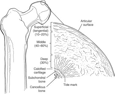
FIGURE 1–3 Articular cartilage layers. (By permission from Brinker MR, Miller MD. Fundamentals of Orthopaedics. Philadelphia: WB Saunders, 1999:9.)
Related posts:
Stay updated, free articles. Join our Telegram channel

Full access? Get Clinical Tree


