Fig. 6.1
(a) Type I denotes fracture of the fifth metatarsal tuberosity extending into the metatarsocuboid joint. (b) Type II describes fracture at the diaphyseal–metaphyseal junction of the fifth metatarsal commonly known as the “Jones fracture.” (c) Type III fractures, also referred to as “proximal diaphyseal stress fractures,” are fifth metatarsal fractures at the diaphysis which is distal to the fourth and fifth intermetatarsal articulation
Type I describes a lateral fracture of the fifth metatarsal tuberosity which extends into the metatarsocuboid joint. Fractures of this area are typically avulsion type injuries resulting from tension from the peroneus brevis tendon and lateral band of the plantar aponeurosis from inversion of the foot.
Type II injuries, known as Jones fractures, involve the metaphyseal–diaphyseal junction of the fifth metatarsal and are a result of indirect adduction mechanism.
Type III injuries describe fractures of the diaphyseal section of the fifth metatarsal which are commonly referred to as proximal diaphyseal stress fractures caused by overuse and repetitive loading.
6.1.4 Distinguishing Between Metaphyseal–Diaphyseal (Jones) and Proximal Diaphyseal Fractures
Fractures of the metaphyseal–diaphyseal junction and proximal diaphysis are common in the athletic population. Because the distinction between the two fractures is not always clear, the term “Jones fracture” has been used interchangeably to describe any fractures proximal to the tuberosity. This is misleading and incorrect. Previous reported series of Jones fractures have revealed inclusion of proximal diaphyseal fractures.6,12–14 Though the anatomic distinction between the two fractures may not always be obvious due to the close proximity of both fractures, differentiation of the two fracture types has been recommended to help guide best possible treatment.
6.1.5 Jones Fractures
In 1902, Sir Robert Jones described a small case series of patients with fifth metatarsal fractures of which the transverse fracture anterior to the metatarsal base bears his name.15 In 1960, Stewart13 later identified a difference in mechanism of injury and prognosis for healing for fractures at the shaft-base junction and fractures of the styloid process. He defined the Jones fracture as a transverse fracture at the diaphyseal–metaphyseal junction that may not extend distal, but through the fourth and fifth metatarsal articulation. The mechanism has been described as adduction to a plantarflexed foot with sufficient force to produce a transverse fracture at the metaphyseal–diaphyseal junction entering the lateral intermetatarsal articulation.12,13 Patients often report pain and mild swelling to the lateral foot and difficulty bearing weight. Since Jones fractures are defined as acute injuries, no prodromal symptoms are reported.8
6.1.6 Proximal Diaphyseal Stress Fractures
Proximal fifth metatarsal fractures, also known as proximal diaphyseal stress fractures, are fatigue injuries primarily seen in the athletic population. The fracture occurs approximately 1.5 cm distal to the tuberosity and is commonly seen as the result of repetitive cyclic stress from high-performance athletic activities.16 This continuous bending stress eventually exceeds the fatigue failure threshold of the metatarsal, leading to microcracks in the bone.17 Prodromal symptoms have been reported in 41% of cases of proximal diaphyseal fifth metatarsal fractures supporting fatigue fracture etiology in these patients.6,7 Criteria for stress fracture of the proximal fifth metatarsal diaphysis has been established by Delee et al.7 to include prodromal symptoms to the lateral foot, radiographic signs of stress fracture, and no prior history of fracture to the fifth metatarsal.
6.1.7 Management of Metaphyseal–Diaphyseal (Jones) and Proximal Diaphyseal Fractures
Recommended treatment for a nondisplaced Jones fracture is immobilization in a non-weight-bearing short leg cast for 6–8 weeks. Surgical intervention is considered for displaced fractures, fractures that demonstrate intramedullary sclerosis or fracture that show little-to-no healing in 3 months. Torg et al.11 reported a 93% healing rate of 15 acute Jones fractures treated in short leg cast non-weight-bearing in an average 6.5 weeks. Clapper et al.8 evaluated 25 Jones fractures treated nonoperatively in a short leg cast non-weight-bearing. They reported 72% healing in an average of 21.2 week with this method. The remaining cohort underwent surgical treatment, 28% achieving healing 12.1 weeks after intramedullary screw placement. Chuckpaiwong et al.18 reported 82.4% healing rate of Jones fractures with nonoperative management in short leg non-weight-bearing cast in 12 weeks and 17.6% developing delayed or nonunion requiring surgical intervention. Porter et al.19 reported 100% clinical healing and 98.9% radiographic healing in 24 surgically treated Jones fractures with a 4.5 mm cannulated screw with return to sports in an average 7.5 weeks.
In contrast, prior reports have suggested prolonged healing periods of up to 21 months and 25% rate of nonunions with proximal diaphyseal stress fractures of the fifth metatarsal with nonoperative treatment.6,20 Therefore, operative management is often recommended for proximal diaphyseal stress fractures. Average radiographic healing rates for operatively managed proximal diaphyseal stress fractures with intramedullary screw range from 6.5 to 9.8 weeks.5,7,18
Chuckpaiwong et al.18 retrospectively compared healing rate of Jones and proximal diaphyseal stress fractures to determine whether distinguishing between the two fractures was necessary to guide treatment. They identified that the overall clinical outcomes of 32 Jones and 29 proximal diaphyseal stress fractures of the fifth metatarsal were not significantly different to warrant differentiating the two fractures. Non-operative care with a short leg cast for at least 4 weeks followed by a short leg cast boot resulted in healing by 12 weeks for up to 87.5% of fractures; however re-fracture and delayed healing was common.
6.1.8 Operative Versus Nonoperative Management of Metaphyseal–Diaphyseal (Jones) and Proximal Diaphyseal Fractures
Though few studies have compared healing rates between operatively versus nonoperatively treated Jones and proximal diaphyseal fractures, operative intervention has shown to have a predictable endpoint of healing in acute and chronic fractures.3,5–8,11–13,16,18,21,22 While little difference is seen in radiographic union rates between operative and nonoperative managed Jones and proximal diaphyseal fractures, operative management offers quicker return to weight-bearing activities and improved patient satisfaction.18 Therefore, operative intervention is recommended for high-performance athletes, recreational athletes, high-demand workers, and displaced fractures to decrease the time to return to sport and high-demand activities. Jones fractures averaged 14.7 weeks quicker return to sport with operative versus nonoperative care (15.3 weeks versus 30.0 weeks). This was a trend also seen in proximal diaphyseal stress fractures which averaged 11.1 weeks quicker return to sports (15.2 weeks versus 26.3 weeks).18
6.1.9 Surgical Technique
Intramedullary screw technique is a minimally invasive method of providing needed compression and stability to these higher risk fractures. The patient is usually positioned in the lateral recumbent position and secured with bean bag positioner (Fig. 6.2). Pillows and blankets are used to pad and position the lower extremity. Fluoroscopic C-arm can then be positioned orthogonal to the operative table facilitating imaging without obstructing the surgical field. A Kirschner wire is placed over the fifth metatarsal to guide axial alignment of the medullary canal and line is drawn with marking pen. The medial border of the fifth metatarsal endosteal cortex or lateral border of the cuboid may be used as guides for axial alignment to the metatarsal (Fig. 6.3). A 2- to 3-cm incision is made proximal to the fifth metatarsal base in line with the skin marking. The sural nerve is often in the proximity of this incision; therefore, due care is exercised to gently retract and avoid nerve injury. Blunt dissection is performed to the fifth metatarsal base. Guide wire for an appropriately diameter cannulated screw (4.0, 4.5, 5.5, or 6.5 mm) and 45–55 mm in length may be used. Alternatively, guide wires may be placed for cannulated drilling; however, often a solid screw is utilized (Fig. 6.4). Starting position of the guide wire is “high and inside” position to ensure axial positioning of the screw23 (Fig. 6.4). Cadaveric study describes the starting position of the guide wire as 1 cm superior to the inferior margin of the tuberosity medial to the peroneus brevis insertion site directed 7° plantarly.24 A more lateral starting position on the fifth metatarsal tuberosity may engage the bow of the metatarsal shaft resulting in gapping of the fracture (Fig. 6.5). Once the guide is past the fracture, appropriately sized cannulated drill is used to drill through the medullary canal. Depth gauge is used to determine appropriate screw length with screw threads passing the fracture site to achieve compression and stability (Figs. 6.6 and 6.7).
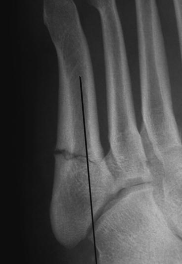
Fig. 6.2
(a, b) Patient is positioned in the lateral recumbent position and secured with bean bag positioner. This position facilitates both surgical access to the fifth metatarsal and ease of fluoroscopic imaging
Fig. 6.3
The medial endosteal border of the fifth metatarsal (denoted with a line) may serve as a guide for axial screw placement into the metatarsal shaft
Fig. 6.4
(a, b) Solid 5.5-mm Jones fracture screw for a Jones fracture
Fig. 6.5
The “high and inside” position on the styloid process of the fifth metatarsal is shown with guide pin (a) and subsequent screw placement (b)
Fig. 6.6
(a) Pre-op proximal fifth stress fracture. (b) A guide wire with a more lateral starting position on the styloid process may be acceptable granted the screw length does not exceed the curvature of the fifth metatarsal which will cause the gapping of the fracture site
Fig. 6.7
Appropriate placement of a well-sized screw will achieve compression and stability of a Jones fracture (a, b) and proximal fifth metatarsal stress fracture (c, d)
6.1.10 Biomechanical Comparisons of Intramedullary Screws
Evaluating the strength of a solid versus cannulated screw, Pietropaoli et al.25 performed three-point bending testing on simulated Jones fractures fixed with intramedullary 4.5 mm malleolar or 4.5 mm partially threaded, cancellous, cannulated screws. Though force at complete displacement was higher for cannulated screw (608.4 N) versus solid screw (519.3 N), there was no significant difference seen between the two screw types in forces at initial or complete displacement.
Sides et al.26 compared bending and pull-out strengths of a 6.5-mm partially threaded lag screw to a variable pitch compression screw with a 4-mm lead thread diameter and 5-mm trailing thread diameter (Acutrak 4/5) screw in simulated Jones fractures. They reported no significant difference in bending stiffness between the two screws but a significant higher resistance to pull-out with 6.5-mm partially threaded lag screw.
Nunley et al.27 compared fatigue resistance of a 4.5-mm thread diameter 3.2-mm core diameter Charlotte Carolina™, Acutrak 4/5, 4.5-mm malleolar, and 4.5-mm cannulated screw to cyclic three-point bending testing. The solid large core diameter Charlotte Carolina™ screw demonstrated significantly higher fatigue resistance in three-point bending compared to the contemporary screws. A limitation of this study was that the method of testing did not replicate physiologic loads that would cause the intramedullary screws to fail.
6.1.11 Postoperative Care
There is little consensus on the postoperative weight-bearing status of surgically treated fifth metatarsal fractures. Management is often conservative non-weight-bearing in a short leg cast for 4–6 weeks. Taking into consideration, high-performance athletes need to return to activities as quickly as possible, most patients are allowed to partial weight-bear as tolerated in a short leg cast in 10–14 days after sutures are removed. Progression into sports-specific training is allowed once pain has resolved and radiographic union is seen. Most athletes return to competitive sports between 7 and 15 weeks after surgery.
6.1.12 Re-fracture
Re-fracture of Jones and proximal diaphyseal fifth metatarsals has been reported in operatively and nonoperatively managed fractures.6,28–30 Re-fracture of proximal diaphyseal fifth metatarsals has been estimated to be less than 5% of these surgically treated fractures.30 The primary risk factor associated with re-fracture of fifth metatarsals was premature return to activity, particularly in elite athletes, prior to radiographic union.29,30 Because re-fracture of the fifth metatarsal is a significant though rare complication, the recommendation is to not remove hardware until the athlete’s professional career is completed. Chronic nonunions (generally from failed nonsurgical treatment) with sclerosis of the fracture margins should be treated surgically. 28–30 These can be treated via a rectangular window, curettement of sclerotic bone, autogenous bone graft insertion, and intramedullary screw placement (Fig. 6.8). The bony window is replaced. Patients are kept in non-weight-bearing status for 6 weeks in a below-knee cast, and then allowed to ambulate with a cast boot for an additional 6 weeks until radiographic union.
Fig. 6.8
(a–d) Jones fracture non-union treated by windowing technique, currettement, autograft and screw placement. (Drawings by Maria Bidny, DPM). (e) Pre-operative X-ray of Jones fracture non-union (Courtesy of Richard Bouche, DPM) (f) Post-operative treatment with dowel-trephine graft from calcaneus with plating (Courtesy of Richard Bouche, DPM)
Related posts:


Stay updated, free articles. Join our Telegram channel

Full access? Get Clinical Tree


