Fig. 12.1
(a) Mortise ankle radiograph in a 35-year-old female recreational runner with 3 months of left ankle pain. The patient was diagnosed with a lateral malleolar stress fracture. Notice a cortical reaction is visible approximately 4 cm proximal to the tip of the lateral malleolus (denoted by arrow). (b) T2-weighted MRI of the same patient. A periosteal reaction in addition to bone marrow edema can be appreciated in the lateral malleolus (denoted by arrow)
In the absence of radiographic findings, advanced imaging with bone scan or MRI is frequently utilized to confirm the diagnosis. MRI has become the advanced imaging modality of choice given improved specificity and ability for concurrent evaluation of the surrounding soft tissues. Periosteal edema, identified on T2 sequence, is the earliest abnormality evident on MRI. With progression of injury, abnormal signal can be detected in the bone marrow on both T1 and T2 sequences, and ultimately progresses to involve the cortical bone (see Fig. 12.1b) [13]. In comparison to patients presenting with normal radiographs, those with evidence of stress fracture on plain films have higher grades of injury as determined with subsequent MRI [13].
Treatment
Nonoperative Treatment
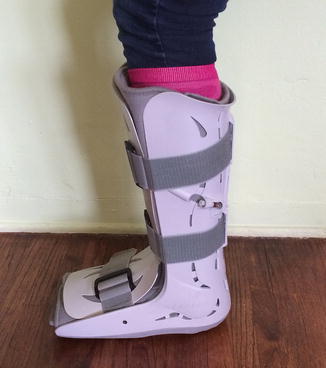
No consensus exists in the literature for nonoperative treatment of fibular stress fractures, but the mainstay of treatment includes rest and activity modification. Early diagnosis is essential in speeding recovery and return to sport. Some patients may benefit from a short period of immobilization with a walking cast or boot to minimize pain and stress at the fracture site; however, the use of an ankle brace, taping, or elastic bandage is preferred over prolonged immobilization [4, 15] (Fig. 12.2) Custom orthotics to correct flexible pes planovalgus or other malalignments may also be beneficial. Full weight bearing is allowed, provided that it does not cause pain, but all high-impact running and jumping activities should be discontinued. During this time, the patient is encouraged to maintain conditioning with low impact activities such as the stationary bike, elliptical trainer, swimming, and pool running. Treatment also incorporates exercises focusing on ankle range of motion, strengthening, and proprioception. Several authors anecdotally suggest that impact activities should be reintroduced gradually when there is no longer tenderness to palpation at the stress fracture site and when compression of the fibula to the tibia does not elicit pain [6, 15]. Most athletes can be expected to return to full activity between 6 and 8 weeks [4, 6, 15].


Fig. 12.2
Example of a walking boot that can be utilized in the nonoperative treatment of stress fractures of the ankle
Operative Treatment
The indications for operative treatment of lateral malleolar stress fractures are limited; however, cases of operatively managed fibular stress fractures do exist in the literature. Kottmeier et al. reported a case of a 19-year-old football player with a non-healing lateral malleolar stress fracture. The patient’s history was notable for recurrent ankle sprains and progressive ossification of the syndesmosis. Resection of the synostosis led to subsequent healing of the stress fracture and return to full activity [22]. Another case study by Guille et al. reported on a patient with an external rotation malunion from a lateral malleolar stress fracture that ultimately required a nonunion take down with bone grafting and open reduction and internal fixation [23].
Stress Fractures of the Medial Malleolus
Incidence, Pathophysiology, and Risk Factors
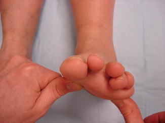
The tibia is the most common site of stress fracture [1, 12, 24]. While 20–60 % of all stress fractures involve the tibial shaft, the incidence of medial malleolar stress fracture is far lower, estimated between 0.6 and 4.1 % [1, 7, 12, 24]. Unlike the low-risk stress fractures of the lateral malleolus, stress fractures of the medial malleolus have limited healing ability, a propensity for displacement, and the potential for prolonged disability. The development of these fractures appears to be highly dependent on repetitive high-impact activity [8, 9, 24–26]. Multiple series report fracture occurrence exclusively in high-level athletes participating in activities requiring significant running and jumping, such as basketball, football, and running [10, 24]. Shelbourne described a cascade of events leading to injury. During the heel strike phase of running, the ankle dorisflexes as the forefoot pronates. With forefoot pronation, the navicular assumes an abducted position relative to the talar head and an internal rotation force is transmitted across the talus. Talar forces are subsequently transferred to the medial malleolus [10]. Repeated high force contact between the talus and medial plafond leads to microtrauma and ultimately stress fracture. Intrinsic factors believed to increase this contact between the talus and medial malleolus include tibia varum and forefoot varus [3, 11, 27] (Fig. 12.3). The presence of an anteromedial talar osteophyte has also been suggested to exacerbate tibiotalar impingement and contribute to the development of fracture [28]. While the majority of these fractures occur in young and middle-aged adults, medial malleolar stress fractures have also been reported in skeletally immature athletes [8, 10, 25, 29].


Fig. 12.3
An example of forefoot varus, a risk factor for stress fracture of the lateral malleolus. Image reprinted with permission of Jeffrey Johnson, M.D.
Presentation
The diagnosis of a medial malleolus stress fracture can be challenging. As such, it is not uncommon for the diagnosis to be delayed weeks to months following the onset of symptoms [9]. Patients often present with vague, ill-defined medial ankle pain in the absence of trauma [8, 10]. As with the fibula, the pain is insidious in nature. It presents initially with activity and then may progress to pain at rest. Some patients may report a rapid exacerbation in pain after a period of mild symptoms. This finding should alert the physician to possible acute displacement of an underlying stress fracture [27]. As mentioned previously, nearly all patients with this injury will report a history of high-impact sports such as endurance running or basketball. They may also report a rapid increase or change in their sporting activities.
Physical Examination
Care must be taken to distinguish this condition from posterior tibial tendonitis and deltoid ligament injury. Physical examination findings include tenderness directly over the medial malleolus at the junction with the tibial plafond [8, 10]. Localized swelling, erythema, and warmth can be present at the site of fracture. Given the intra-articular nature of the fracture, an effusion of the ankle joint may be present and limit ankle range of motion [7, 10]. Running and/or jumping activity will often reproduce the patient’s pain [10]. As mentioned previously, a thorough examination of standing alignment and gait must be performed and compared to the contralateral side.
Imaging
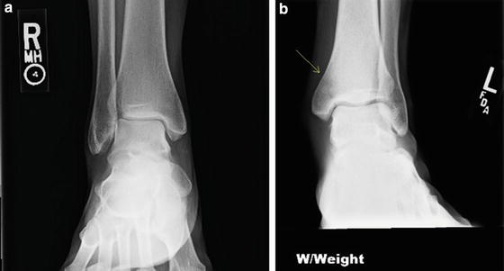
The high proportion of cancellous bone in the medial malleolus makes detection of a stress fracture difficult, even after the early phase of healing (4–6 weeks) when most other stress fractures become visible on plain radiographs (Fig. 12.4a). Early osseous changes, when present, include blurring of the trabecular margins and subtle sclerosis from peritrabecular callus formation. Over time a sclerotic line may be apparent within the cancellous bone. The characteristic medial malleolar stress fracture line appears on the posteromedial, concave side of the tibia and extends in a vertical or oblique direction from the junction of the medial malleolus and the tibial plafond into the tibial metaphysis (see Fig. 12.4b) [8, 10, 25]. Small lytic lesions surrounding the fracture line have also been described [25].


Fig. 12.4
(a) AP ankle radiograph of a 47-year-old active female with 2 weeks of medial ankle pain. These radiographs are normal but the patient went on to be diagnosed with a medial malleolar stress fracture. (b) AP radiograph of a 32-year-old male professional athlete with a clearly visible stress fracture line in the medial malleolus
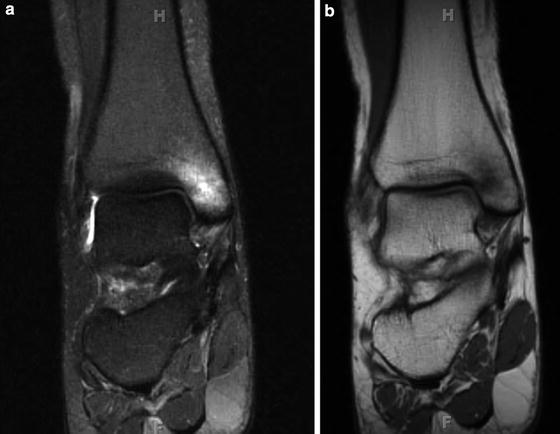
In some cases, changes on plain radiographs may never become visible [9]. Advanced imaging is recommended in all patients with negative radiographs, but with clinical signs and symptoms concerning for a stress fracture a high index of suspicion should be employed [9]. As with the fibula, MRI is preferred for the early detection of medial malleolar stress fractures [9]. The earliest osseous changes can be depicted on fat suppressed T2-weighted images or STIR sequences where soft tissue and bone edema result in high signal changes within the cancellous bone (Fig. 12.5a). As the injury and associated edema progress, abnormalities become visible on T1-weighted images as a linear area of low signal that runs perpendicular to the trabeculae and ultimately extends into the cortex (see Fig. 12.5b) [19]. Recently, ultrasound has also been suggested as a possible imaging modality for the diagnosis of medial malleolar stress fractures [21]. In a small series of patients, periosteal thickening, cortical irregularities, edema and pain with transducer compression were appreciated in nearly all patients. Furthermore, another recent study proved that ultrasound was 83 % sensitive and 76 % specific for metatarsal stress fractures when compared with MRI [30]. This low-cost modality could become more popular and beneficial in diagnosing malleolar stress fracture in the future; however, as with all ultrasounds imaging, the results are highly dependent on the skill of the ultrasound technician performing the exam. Bone scan may also be used in diagnosing medial malleolar stress fractures, but this imaging technique has been largely supplanted by MRI.


