Fig. 9.1
The intra-articular disk ligament (Held by forceps), costoclavicular ligament (Figure B arrowhead), capsular ligament (Figure B arrow), and interclavicular ligament lend integrity to the sternoclavicular joint
Mechanism of Injury
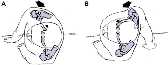
Due to the strong ligamentous supporting structure of the sternoclavicular joint, it requires tremendous force to dislocate the joint. These forces may be applied via direct or indirect application to the shoulder. Direct forces to the sternoclavicular joint typically result in posterior forces to the joint. Indirect forces (Fig. 9.2) are the most common mechanism of injury and may result in either anterior or posterior direction of forces across the sternoclavicular joint [4, 5]. Motor vehicular accidents and athletics account for over 80% of the cases of injury to this joint [6–8].


Fig. 9.2
(a) Forces resulting in an anterior dislocation of the sternoclavicular joint. (b) Forces resulting in a posterior dislocation of the sternoclavicular joint
Classification of Injury
Sternoclavicular joint subluxation/dislocation injuries are uncommon. They can be organized by degree (subluxation, dislocation), timing (acute, chronic, recurrent), direction (anterior, posterior), and cause (traumatic, atraumatic).
Atraumatic Subluxation and Dislocation
Atraumatic SCJ dislocation/subluxation is an uncommon injury which occurs most often in adolescent female overhead athletes with multijoint laxity. On the basis of its bony anatomy, the SCJ is inherently unstable. However, the strong costoclavicular and intraclavicular ligaments, as well as capsular support, render the SCJ extremely stable. Atraumatic SCJ subluxation/dislocation must be distinguished from traumatic injury, as the treatment strategies and outcomes are significantly different [9].
Physical examination typically reveals that the patient to be neurovascularly intact, with symmetric shoulder girdle appearance without any visible abnormalities, and full shoulder range of motion bilaterally. Active and passive abduction with forward flexion may provoke SCJ subluxation which was accompanied by an audible click. The shoulder examination also typically reveals glenohumeral joint multidirectional instability, with hyperlaxity also present at wrist, elbow, finger, knee, and ankle joints.
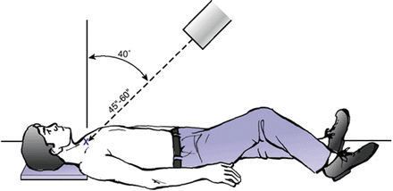
Radiographic evaluation of these cases includes standard three view radiographic examinations of the shoulder (ap, axillary lateral and scapular y views). In an office setting, the addition of a serendipity view as described by Rockwood [10] (Fig. 9.3) may be beneficial. Further radiographic studies should typically include a computerized tomogram (Fig. 9.4a, b). If there is a question of physeal injury, an MRI examination can separate the difference between physeal injury and subluxation/dislocation.


Fig. 9.3
Serendipity view may be beneficial

Fig. 9.4
(a) Computerized tomogram demonstrating a posterior dislocation. (b) 3 Dimensional image of a posterior sternoclavicular dislocation
The differential diagnosis includes atraumatic subluxing SCJ, secondary to congenital multijoint hypermobility. Other diagnostic considerations to exclude would be ligamentous and bony abnormalities, including the growth plate, and degenerative or neoplastic lesions.
Treatment
Most patients seek medical treatment because of initial pain and concern regarding the potential harm of the condition. In a review of 37 patients with spontaneous anterior subluxation of the sternoclavicular joint [11], subluxations were reproducible and painless in 29 patients. Eighty percent of the patients demonstrated evidence of generalized ligamentous laxity. Twenty-nine patients were treated nonsurgically with strengthening exercises and advancement to unrestricted activity as tolerated. Although many patients subsequently reported intermittent episodes, few reported discomfort, and most were able to participate successfully in athletics.
The most common reason for surgery was the failure of a previous attempt at reconstruction. Surgery is rarely indicated. Nonsurgical management, including patient education of the benign nature of the condition, is recommended [11]. For patients with continued symptoms in spite of physical therapy, sternoclavicular joint reconstruction with a figure-of-eight graft may be indicated. Short- and intermediate-term clinical results of this reconstruction [12] have shown promise. However, longer term studies are currently lacking regarding the durability of results. The surgical technique is described in the following section outlining anterior SC dislocation.
Strains and Subluxations
In a mild sprain , the ligaments of the sternoclavicular joint remain intact. The patient reports pain and tenderness with palpation over the joint. Swelling may be present, but no instability is noted. Swelling and pain become more pronounced as the ligaments are stretched, which results in subluxation of the joint. Pain is marked with motion of the ipsilateral extremity. Laxity of the joint may be apparent compared with the contralateral joint.
Dislocation
Severe pain and deformity accompany dislocations of the sternoclavicular joint. Surprisingly, clinical examination to determine the direction of the dislocation may be inconclusive because of swelling. In addition to swelling, compression of the vital structures posterior to the joint may occur with dislocation injuries. The orthopedic surgeon should keep this potential life-threatening complication in mind while performing the clinical evaluation.
Anterior sternoclavicular injuries may exhibit prominence of the medial clavicle. This prominence is more easily appreciated while the patient is in the supine position. Posterior dislocation is less common than anterior dislocation. Patients with posterior dislocation demonstrate a higher level of pain, and the corner of the sternum may be discerned as the medial clavicle is displaced posteriorly [7]. However, swelling may preclude an accurate clinical assessment of injury.
Patients with posterior displacement may report shortness of breath or difficulty breathing Because of compression of the trachea or pneumothorax. Similarly, compression of the esophagus may result in dysphagia. Compression of the posterior vascular structures can result in decreased circulation to the ipsilateral extremity or venous congestion in the extremity or neck. Tingling or numbness may be the predominant complaint with compression of the brachial plexus. Posterior sternoclavicular dislocation or associated injuries may render the patient medically unstable.
Radiographic Evaluation
Routine radiographic studies of the sternoclavicular joint are difficult to interpret. Rockwood [10] developed an oblique view of the sternoclavicular joint dubbed the serendipity view. This radiograph is obtained by pointing the radiographic beam at a 40° angle tilted cephalic with the beam centered on the sternoclavicular joint. The resulting radiograph allows for comparison of the relationship of the injured clavicle to the normal clavicle. The technique is best suited for isolated sternoclavicular injuries.
Plain radiographic examination and oblique views have largely been supplanted with the availability of computerized tomograms. CT scanning is far superior technique over conventional radiographs to study any problems of the sternoclavicular joint (Fig. 9.3). These scans easily distinguish sprains, dislocations, and medial clavicle fractures. In order to assist the radiologist, information pertaining to the history and mechanism of injury is provided. An accurate diagnosis is further facilitated by ensuring that the scan includes both medial clavicles so that the injured sternoclavicular joint can be compared to the opposite side. The examination should also include a CT of the chest to identify any associated injuries.
In children and young adults, MRI is especially helpful in distinguishing a dislocation of the sternoclavicular joint from a physeal injury. The ability of MRI to image soft tissue also allows assessment of the integrity of the costoclavicular ligament and attachments of the intra-articular disk. Imaging and location of the trachea, esophagus and great vessels are also available with MRI. CT examination is the imaging medium of choice in the acute situation due to its speed, availability, and ability to image bone injury.
Treatment
Anterior Strain/Subluxation
The utilization of ice and analgesics in the initial treatment of these injuries is advocated. Subluxations may be reduced by directing the shoulders posterior and medial. Support of the injury by utilization of a clavicle strap or sling and swath is typically useful. The patient is protected from injury using immobilization for 6 weeks after injury.
Anterior Dislocation
Closed reduction of anterior dislocation is the current treatment of choice although there is still some controversy regarding management [13, 14]. Closed reduction may be accomplished with sedation, local or general anesthesia. The patient is placed supine on a table with a 3 in. pad between the shoulders. Pressure is placed posteriorly on the medial clavicle. If the joint remains reduced, the patient is immobilized (figure-eight or velpeau type sling) for 6 weeks is utilized to allow healing.
Unfortunately, most anterior sternoclavicular dislocations are unstable after closed reduction, but if successful results in improved cosmesis. Anterior dislocations frequently will remain chronically unstable but generally do not deteriorate to debilitative symptoms [11, 15]. Good results have been shown with nonoperative treatment of asymptomatic anterior instability by physical therapy and activity modification. In rare instances, patients may develop painful crepitus with arm motion and pain that radiates into the neck, altering their ability to work and to participate in athletics [16, 17].
Although numerous methods of open reduction have been described [18–20], most injuries are asymptomatic. Surgical treatment remains controversial as indications and techniques continue to evolve. In 2004, Spencer and Kuhn [21] reported a biomechanical study investigating the strength of a reconstructed sternoclavicular joint utilizing a figure-of-eight semi-tendinosis graft which demonstrated superior biomechanical properties. Multiple clinical studies have demonstrated good results utilizing the technique [22] with minor technical changes incorporated since the initial report including utilization of suture anchors and allograft tissue. Sabatini et al. [12] reported the most recent results of this technique in ten patients with significant improvements in pain scores and ASES scores with a minimum of complications.
Surgical Technique
The exposure for this technique reported by Sabatini et al. [12] is similar to what has been previously described [23–25] (Fig. 9.5). Briefly, the patient was placed in a supine position and prepared and draped (left shoulder and anterior chest), with the assistance of a cardiothoracic surgeon; a slightly angled transverse incision was made from the medial aspect of the clavicle to the manubrium. This area was then dissected, and the platysma was incised and raised, followed by elevation of periosteum off of the clavicle. The SCJ was visualized and the capsule reflected superiorly and inferiorly, providing room for the reconstruction.


Fig. 9.5
(a) Drilling holes in clavicle and sternum for the reconstruction. (b) Passing tendon reconstruction through holes. (c) Reconstruction is secured in a figure eight technique
Typically, it is necessary to elevate the soft tissues surrounding the manubrium with removal of the intra-articular disk. Once the approach had been completed and adequate deep visualization of the manubrium and the clavicle was obtained, the allograft was prepared. Typically, a 4.5-mm 25-cm semitendinosus allograft was used with No. 2 polyethylene suture on each end. Four drill holes were placed by a 4.5-mm drill with a cannulated system, 2 in the manubrium and 2 in the distal clavicle.
After this, the graft was shuttled in the sequential fashion from the inferior manubrial drill hole to the inferior clavicle back to the superior manubrial drill hole back to the superior clavicle drill hole.27 With the graft tensioned, two 4.75-mm-diameter 15-mm-length PEEK tenodesis screws (Arthrex, Naples FL, USA) were then placed within the first (inferior manubrium) and fourth (superior clavicle) drill holes to secure the graft. After the placement of the tenodesis screws, two figure-of-eight stitches were placed with No. 2 polyethylene suture to secure the two sides of the graft to one another for additional support without the prominence of a knot of tied tendon. The suture knots were subsequently buried. The sternocleidomastoid, which was initially taken down, was then reapproximated with No. 1 Vicryl. The subcuticular layer was closed with 2-0 Vicryl (Ethicon, Somerville, NJ, USA), and then the skin with staples.
Postoperatively the patient is immobilized in a sling and allowed elbow and hand range of motion immediately postoperatively. Pendulum exercises are instituted within the first 2 weeks after surgery and passive range of motion exercises for the shoulder are continued through the sixth week after surgery. At this point, a gentle strengthening program is instituted and the sling may be discontinued. Activity restriction is typically enforced until 16 weeks after surgery and a return to sport may occur at 6 months postoperatively.
Posterior Dislocation
A careful history and physical examination of these injuries and a low threshold for the utilization of CT imaging is recommended. If evaluation reveals dyspnea, choking, or hoarseness, this indicates pressure on the mediastinum. Mediastinal involvement requires prompt consultation with a thoracic or cardiothoracic surgeon in the management of these individuals.
Closed Reduction
Open techniques are typically not required to reduce acute posterior sternoclavicular injuries. Further, once the reduction is achieved via closed reduction it is typically stable [26]. The authors have a thoracic surgeon available during closed reductions.
Technique
Under sedation or general anesthesia , the patient is placed supine on the operating room table. Folded towels or sandbags, 3–4 in. thick, are placed between the scapulae to extend the shoulders. The involved extremity is positioned near the edge of the table, allowing the arm to be extended and abducted (Fig. 9.6). Initially, gentle traction is applied to the abducted extremity in line with the clavicle while counter traction is applied by an assistant who steadies the patient. The traction on the arm is slowly increased while the arm is brought into extension. If this reduction technique fails [27], traction may be applied to the arm in adduction while a posterior pressure is applied to the shoulder in order to lever the clavicle over the first rib.


Fig. 9.6
Closed reduction manuevers for treatment of a posterior sternoclavicular dislocation include positioning the extremity near the edge of the table, allowing the arm to be extended and abducted
Stay updated, free articles. Join our Telegram channel

Full access? Get Clinical Tree


