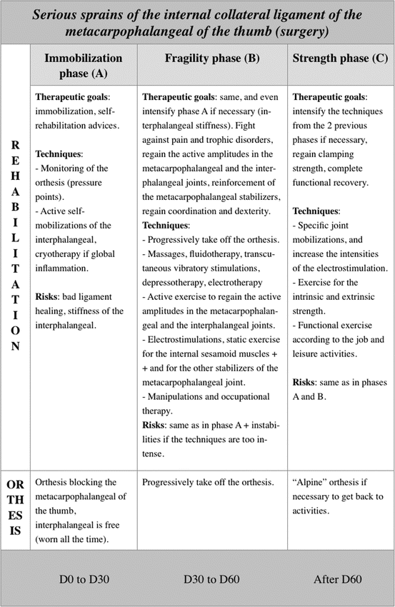
Fig. 6.1
The automatic rotation of the metacarpophalangeal of the thumb in pronation during the opposition between the thumb and the finger is related to ligamentous (ulnar collateral ligament shorter than radial collateral ligament), muscular (external thenar muscles more active), and osseous (external part of the first metacarpal head longer than the internal one) factors
The passive stability of the joint is mainly ensured by the collateral ligaments and the volar plate (Fig. 6.2).


Fig. 6.2
Ligamentous system of the metacarpophalangeal of the thumb
The collateral ligaments are made of two fascicles:
The principal fascicle goes from the head of the first metacarpal to the base of the first phalanx, slightly dorsal to the joint axis in flexion/extension. In flexion, it’s tensed, ensuring the lateral stability.
The accessory fascicle is oblique at the palmar and distal level; it goes from the head of the first metacarpal to the corresponding sesamoid, thus creating a common structure with the volar plate. It’s tensed in extension, ensuring the lateral stability in this position.
The volar plate is a thick and resistant fibrocartilage, which contains the two sesamoid bones and opposes the metacarpophalangeal extension.
The most instable joint position is the intermediate flexion, as the collateral ligaments and the volar plate are relaxed.
The active joint stability is ensured by the internal (adductor pollicis, inconstant first palmar interosseous) and external (abductor brevis and flexor brevis) sesamoid muscles (Fig. 6.3).
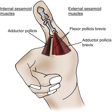

Fig. 6.3
Active stability of the metacarpophalangeal joint of the thumb, depending mainly on the internal and external sesamoid muscles
The combined movement of flexion–radial inclination–pronation made by the external and internal thenar muscles allows optimizing the cylindrical grips with a full palm, increasing the contact surface.
The opponens metacarpal pollicis, which ends on the palmar side of the first metacarpal, doesn’t play a role in the stability of the metacarpophalangeal joint, and the extrinsic muscles only have a moderate action.
6.2 Physiopathology [2]
The metacarpophalangeal sprains are frequent in the thumb due to sports (skiing, handball, basketball, rugby, etc.) [3, 4] and are the results of injuries in the valgus in 90 % of the cases (damage of the medial collateral ligament).
The traumas in the varus damage the lateral collateral ligament, and the ones in hyperextension damage the volar plate. Injuries with a combination of various elements often occur.
In serious sprains of the medial collateral ligament (rupture), ligament healing is impossible and surgery is needed [5, 6], as the proximal extremity of the ligament passes over the adductor’s expansion toward the extensor pollicis longus, making the healing impossible: it’s the Stener effect (Fig. 6.4).
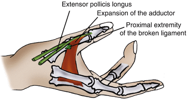

Fig. 6.4
Physiological explanation of the Stener effect
Even if it’s rarer, the same phenomenon can be observed radially, with the expansion of the external thenar muscles.
The varus deformity in the injuries of the radial collateral ligament is more spectacular than the valgus deformity in the injuries of the ulnar collateral ligament [7], due to the longitudinal orientation of the external thenar muscles and the transversal orientation of the adductor pollicis, which will have an important tendency to lead to varus in case of an injury of the radial collateral ligament (Fig. 6.5).
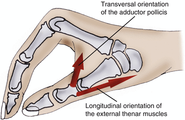

Fig. 6.5
The varus deformity in case of an injury of the radial collateral ligament is important because the adductor pollicis is transversal to the osseous lever, unlike the external sesamoid muscles
6.3 Clinical and Paraclinical Signs
6.3.1 Clinical Signs
Other than the usual signs of a damaged ligament (pain when palpating and when putting it in tension, swelling, sensation of instability), we have to be careful to some specific clinical signs that allow suspecting a serious sprain with a Stener effect:
Hematoma on the dorsal part of the interphalangeal joint (pathognomonic)
Valgus 30 % superior to the contralateral side and metacarpophalangeal joint flexed to 30° (specific test of the metacarpophalangeal fascicle) (Fig. 6.6) [7]
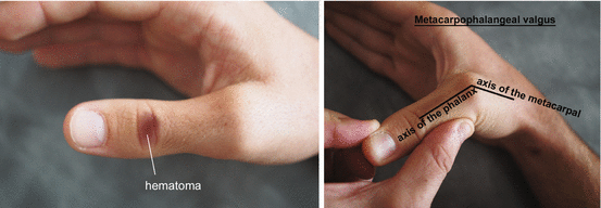
Fig. 6.6
Clinical signs leading to suspecting a Stener effect; hematoma on the dorsal side of the interphalangeal (left) and valgus 30 % superior to the contralateral side (right)
6.3.2 Paraclinical Signs
The bilateral test in valgus, under a radiological control, allows obtaining a better appreciation of the angular value than the simple clinical test.
The presence of a bone fragment at the level of the phalangeal insertion means there is a rupture of the ligament, with a Stener effect.
6.4 Therapeutic Process and Surgical Treatment [5, 6, 8, 9]
In the case of a benign sprain (ligamentous distension without important slackness or osseous avulsion), we suggest an immobilization of the metacarpophalangeal joint in a functional position (without any varus/valgus component), leaving the wrist and the interphalangeal joint free.
The Orthosis is realized in a thin plastic (1.6 mm) and kept for 3–4 weeks to allow ligamentous healing.
In the case of a serious sprain with a Stener effect (important slackness with or without osseous avulsion), the treatment is surgical.
It consists in reinserting the ligament at the level of the first phalanx, with an intraosseous anchor, in order to regain good stability of the metacarpophalangeal (Fig. 6.7).
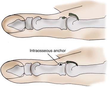

Fig. 6.7
Intraosseous anchor of the ligament in case of a serious sprain with Stener effect
A postoperative Orthosis is realized with the same criteria as the one made for benign sprains. It’s kept for 4 weeks.
6.5 Rehabilitation and Orthosis
The metacarpophalangeal joint of the thumb is a stabilizing joint. The aim of the rehabilitation is to improve active stability of the joint, particularly the muscles protecting the damaged ligament. The techniques to gain amplitude will be used very carefully during the healing period, in order not to weaken the passive stabilization structures.
In case of surgery, ultrasounds mustn’t be used if a metallic intraosseous anchor has been used.
6.5.1 Rehabilitation Protocol of the Serious Medial Collateral Ligament Sprains (Postoperative) (Fig. 6.8)

