Figure 11.1
Lateral radiograph demonstrating L5/S1 spondylolysis
1.
Grade I—0–25% slip.
2.
Grade II—26–50% slip.
3.
Grade III—51–75% slip.
4.
Grade IV—76–99% slip.
5.
Grade V—Spondyloptosis, or 100% slip.
The presentation is with lower back pain, which is usually unilateral, and worse with hyperextension or rotation. Examination reveals hyperlordosis, weak abdominal muscles and tenderness that is worst at the belt line. Symptoms are reproduced by extension, particularly unilateral extension when the patient leans back whilst standing on one leg. Pain is worst on the ipsilateral side. There may also be relative hamstring tightness and a pelvic waddling gait. Normal neurology can be expected for pars defects, but with higher grades of spondylolisthesis, signs of compression may be present.
Initial imaging with plain radiographs including AP, standing lateral and both oblique views, can show the pars defect “collar” on Lachapele’s “Scotty dog” view. As the athlete may present very early, a technetium 99 bone scan may highlight a defect before radiographic findings. They are also of benefit in differentiating acute lesions needing more aggressive treatment, from chronic lesions. SPECT (single photon emission computed tomography) and CT (computed tomography) add further information to guide treatment.
Management of acute spondylolysis varies, but the consensus involves short periods of bed rest if required, or the utilization of a brace, followed by a generalized fitness program where aggravating activities are avoided. Treatment of chronic lesions is symptomatic. Spondylolisthesis, if less than 50%, may be managed non-operatively and sport may be allowed, albeit with strong advice regarding potential of further slippage and long term problems. If more than 50%, restriction of activities is recommended and surgery may be considered. For further information, see Chap. 5.
11.6 Shoulder Injuries
11.6.1 Anterior Shoulder Instability
Anterior shoulder instability related to sport is associated with the combination of abduction, external rotation and extension of the joint. The spectrum includes acute dislocations, recurrent subluxations as well as recurrent dislocations. Clinical presentation is with pain, a tender anterior swelling, and “squaring-off” of the shoulder. Examination characteristically demonstrates absence of external rotation whilst the shoulder is dislocated, and must include neurovascular assessment pre-reduction, especially for the axillary and musculocutaneous nerves. Plain radiographs including anteroposterior and Y- or axillary views can confirm or exclude an associated fracture.
Management consists of reduction, immobilization and rehabilitation. Reduction can be done via one of several methods, with aid of muscle relaxants and anaesthesia when available. Care must be taken not to cause traction injury to nerves. Scapular rotation or Stimson’s methods are done with the patient prone, whilst the modified Kocher’s, double sheet and Hippocratic methods are utilized when supine. Neurovascular examination and radiographs must be repeated post-reduction to ensure that the humeral head is located and no iatrogenic complications have occurred. Immobilisation to allow for capsular healing should be followed by graded range of movement and muscle strengthening exercises. Return to sport should be delayed for at least 3 months.
Athletes may often have an increased range of movement than normal, particularly those involved in throwing or swimming. This asymptomatic movement may border on subluxation. Recurrent subluxation may also cause these athletes to present with pain mimicking impingement, clicking or neurological symptoms. Recurrent dislocation may have a voluntary element, which may require psychiatric input. Surgical stabilization in voluntary dislocators is more prone to failure, so this must be assessed prior to any operation. Examination of range of movement and stability includes the anterior apprehension test and observing for the sulcus sign. Examination under anaesthesia may also be of diagnostic benefit. Note should be made of the patient’s general laxity, e.g. using Beighton’s score. Imaging may show a Hill Sachs or Bankart lesion. Operative procedures include the open Bankart procedure, with or without capsular shift, and arthroscopic stabilization.
11.6.2 Proximal Humeral Epiphysiolysis (Little League Shoulder)
Repetitive throwing can cause chronic microtrauma to the proximal humeral epiphysis. Patients present with limitation of activity and localized tenderness on palpation. Widening of the epiphysis can be seen on radiographs. Treatment is symptomatic, with rest until comfort allows return to throwing.
Clavicular and proximal humeral fractures, and acromioclavicular joint injuries are covered in Chap. 2.
11.7 Elbow Injuries
11.7.1 Little League Elbow
Young throwing athletes are susceptible to chronic valgus overloading, resulting in repetitive microtrauma to the medial and lateral structures, varying with the phases involved in the throwing motion and the patient’s age. Medial pain results from valgus traction causing medial epicondyle apophysitis, avulsion fractures, medial epicondylitis and ulnar collateral ligament sprains or tears, with increasing skeletal maturity. Laterally, compressive forces acting on the capitellum during cocking and acceleration may lead to avascular necrosis (Panner’s disease) or osteochondritis dissecans. Conversely, traction forces in the follow-through phase produce apophysitis of the lateral epicondyle or injury to the radial physis.
Clinical presentation is with elbow pain, predominantly affecting the medial side that impairs the athlete’s performance. Radiographs may be normal, or indicate particular age-related pathology. Imaging of the non-affected side together with stress views may be of benefit. MRI may have increased sensitivity, but clinical suspicion must prompt treatment. Management is by resting the elbow from the causal stresses for 4–6 weeks. Following this, a graded return to throwing over a further 4–6 weeks may be started.
11.8 Hip and Pelvic Injuries
11.8.1 Avulsion Fractures and Apophysitis
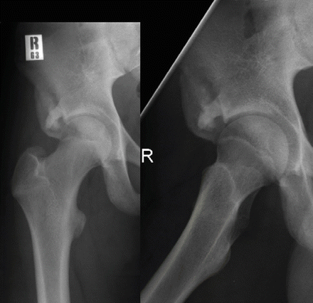
Avulsion fractures occur rather than strains, particularly at the origins or insertions of muscles around the pelvis or hip. The mechanism may be by indirect trauma or chronic repetitive microtrauma. Areas typically affected include the anterior superior iliac spine (ASIS) due to the pull of sartorius, the anterior inferior iliac spine (AIIS) due to rectus femoris (Fig. 11.2), the ischial apophysis due to the hamstrings, and the lesser trochanter due to iliopsoas. Radiographs help confirm the diagnosis and are useful in follow-up, when the secondary ossification centre can be seen displaced from its anatomical location. Confusion may arise when the secondary ossification centre is not yet visible. Management is usually non-operative, with limitation of muscle usage followed by graded return to sport. Surgical repair is usually reserved for large fragments or resultant loss of function, which is more common at the elbow’s medial epicondyle and the knee’s tibial tubercule. There are usually no long-term limitations, unless the resultant healed bone causes secondary impingement.


Figure 11.2
Chronic pincer changes following an anterior inferior iliac spine avulsion
11.8.2 Iliac Crest Apophysitis
Iliac crest apophysitis usually affects the anterior half of the crest and is caused by pull of the abdominal wall musculature. It is seen in the adolescent runner complaining of persistent hip pain limiting participation. Examination reveals tenderness over the crest and pain on lateral rotation. Management is by modification of activity including avoidance of running for several weeks whilst maintaining fitness through other exercises.
Stay updated, free articles. Join our Telegram channel

Full access? Get Clinical Tree


