Age
AP-BMD
L-BMD
Mid-BMD
FN-BMD
TR-BMD
WD-BMD
Osteophyte score
Disc score
No. of vertebral fractures
Age
−0.218*
−0.306**
−0.254*
−0.327**
−0.328**
−0.291*
−0.027
0.016
0.295**
AP-BMD
0.727***
0.669***
0.740***
0.590***
0.656***
0.580***
0.570***
−0.395***
L-BMD
0.912***
0.526***
0.464***
0.523***
0.349***
0.480***
−0.390***
Mid-BMD
0.445***
0.400***
0.462***
0.344***
0.445***
−0.347***
FN-BMD
0.847***
0.806***
0.417***
0.340**
−0.524***
TR-BMD
0.765***
0.330**
0.233*
−0.406***
WD-BMD
0.263*
0.298*
−0.466***
Osteophyte score
0.741***
−0.152
Disc score
−0.266**
No. of vertebral fractures
The effects of spondylosis on increasing BMD in the lumbar spine have been emphasized [16, 46, 50]. However, the methodology of our study eliminated the substantial contribution of osteophyte formation and disc degeneration to the BMD measurement, because higher BMD in Ward’s triangle, which is a subregion primarily consisting of the trabecular bone, and higher BMD in the proximal femur, which is a site remote from the lumbar spine, were observed in patients with higher osteophyte scores and disc scores.
7.2 Pharmacotherapy for Osteoporosis and the Effect of Coexistent Spondylosis
7.2.1 Anti-Vertebral Fracture Effect of Spondylosis
Since osteoporosis and spondylosis often occur simultaneously, treatment of osteoporosis with antiosteoporotic agents may be affected by the coexistence of spondylosis in many patients. The presence of spondylosis may have a preventive effect against vertebral fractures. However, there are few reports about the effects of osteoporosis medication on the incidence of vertebral fractures in people with spondylosis.
We previously conducted a retrospective investigation of the effects of alfacalcidol alone or in combination with elcatonin on the incidence of osteoporotic vertebral fractures in women with spondylosis [34]. The severity of spondylosis was evaluated with the aforementioned “osteophyte score” [32]. The study subjects were 101 postmenopausal women with osteoporosis aged >60 years, divided into three groups: the D group (n = 45), treated for >5 years with alfacalcidol; the D + ECT group (n = 26), treated for >5 years with alfacalcidol plus elcatonin ; and the control group (n = 30), received no medications for >5 years. Over the 5-year treatment period, the number of incident vertebral fractures per patient was significantly higher in the control group (2.9) than in the D group (1.2) and D + ECT group (1.5) (p < 0.01) [34]. However, in all three groups, the number of incident vertebral fractures was positively correlated with the number of prevalent vertebral fractures (0.303 ≤ r ≤ 0.434), and it was negatively correlated with baseline BMD (−0.703 ≤ r ≤ −0.329) and the osteophyte score (−0.769 ≤ r ≤ −0.365) [34]. Further multiple regression analysis showed that pharmacotherapy (D or D + ECT, p < 0.001) and the osteophyte score (p < 0.001) were the most significant contributors to the number of incident vertebral fractures [34]. These results showed that the presence of spondylosis (indicated by a high osteophyte score) appears to have an effect on the prevention of vertebral fractures. Because this study was designed as a retrospective study, for this chapter, we further evaluated the anti-vertebral fracture efficacy of pharmacotherapy and spondylosis in a prospective, randomized study described below.
7.2.2 Effect of Spondylosis on the Results of Early-Phase Osteoporosis Treatment with Alendronate and Alfacalcidol: A 6-Month, Prospective, Randomized Study
7.2.2.1 Background
Bisphosphonates are the most popular antiosteoporotic agents and are used worldwide. However, because the effects on the bone are exerted in an indirect manner by reducing the remodeling space and prolonging the duration of mineralization, several months are required to increase bone mass and strength. Previous reports have shown that significant antifracture effects of alendronate can be expected after 6 months of treatment [24]. Alfacalcidol also shows preventive effects against osteoporotic fractures, despite a small effect on bone mass [45]. We thus conducted a 6-month, prospective, randomized trial of postmenopausal women with osteoporosis to evaluate the possibility of early-phase superiority using combined treatment with alendronate and alfacalcidol compared to either alone, with radiographically diagnosed vertebral fracture as the primary end point. The preliminary results related only to fracture incidence were reported in Japanese and showed that combination therapy with alendronate and alfacalcidol was superior in terms of preventing vertebral fractures over either treatment alone in early-phase treatment (≤6 months) [33]. However, because many of the enrolled osteoporotic patients also had spondylosis, the antifracture efficacy of spondylosis might also have affected the results. In addition, several other risk factors, such as spinal hyperkyphosis, lower BMD, and higher bone turnover markers, were not included in the preliminary report. Because we have many unpublished data from this study, we reevaluated the anti-vertebral fracture efficacy of alendronate and alfacalcidol in the presence of spondylosis for this chapter.
7.2.2.2 Methods
A total of 441 Japanese women with postmenopausal osteoporosis aged 60 years and over who initially attended one of two institutions (Minamiakita Orthopedic Clinic, Katagami, Japan, and Igarashi Memorial Hospital, Akita, Japan) and showed interest in participating in this study was enrolled. Osteoporosis was diagnosed according to the year 2000 version of the Diagnostic Criteria for Primary Osteoporosis published by the Japanese Society for Bone and Mineral Research [44]. The exclusion criteria were as follows: (1) women with a history of metabolic bone disease except for postmenopausal osteoporosis, malignancy, or previous antiosteoporotic treatment, (2) chronic glucocorticoid usage, and (3) patients with documented vertebral and/or nonvertebral fractures within the last 6 months.
The patients were randomized to one of three groups: ALN group, treated with daily oral administration of 5 mg of alendronate (Bonalon; Teijin Pharma, Tokyo, Japan); D group, treated with daily oral administration of 1 μg of alfacalcidol (Alfarol; Chugai Pharmaceutical, Tokyo, Japan); and ALN + D group, treated with daily oral administration of 5 mg of alendronate plus 1 μg of alfacalcidol . This study was conducted with a prospective, randomized, open-label design, with a duration of 6 months. Spinal X-rays, BMD of the distal radius, and serum samples for bone turnover markers were obtained at baseline and final follow-up (at 6 months). This study was performed in accordance with the recommendations of the Declaration of Helsinki, and patients’ informed consent was obtained before randomization.
Thoracic and lumbar spine X-rays with anteroposterior and lateral views in a neutral position were taken using a film-tube distance of 1.0 m [32]. The thoracic radiographs were centered at T8 and the lumbar radiographs at L3 [32]. Vertebral fracture was considered present if at least one of three height measurements (anterior, middle, and posterior) for one vertebra had decreased by >20 % compared with the height of the nearest uncompressed vertebral body [45]. Angles of kyphosis for the thoracic (T4–T12) and lumbar (L1–L5) spines were also measured from lateral radiography using the Cobb angle method. As an indicator of spondylosis, osteophyte formation was assessed at baseline using the semiquantitative osteophyte score as mentioned above [32]. All radiographic assessments were made by two expert spine surgeons (NM, YK) masked to the BMD values and treatment groups.
BMD was measured at the distal 1/3 radius by DXA (DTX-200; Toyo Medic, Tokyo, Japan). Serum samples were obtained before noon after at least a 3-h fast, and N-terminal telopeptide of type I collagen (NTX) and bone-specific alkaline phosphatase (BAP) were measured at baseline and 6 months after the beginning of the treatment. Serum NTX was measured by an enzyme-linked immunosorbent assay (Osteomark; Mochida Pharmaceutical, Tokyo, Japan; reference range 9.5–17.7 nmolBCE/L) as a marker of bone resorption. Serum BAP was measured with an enzyme immunoassay kit (Osteolinks-BAP; Sumitomo Pharmaceuticals, Tokyo, Japan; reference range 13.0–33.9 U/L) as a marker of bone formation.
Statistical analysis of differences among the three groups was performed using Fisher’s protected least significant difference method (a post hoc test) for multiple comparisons in one-way analysis of variance (ANOVA). A paired or unpaired t-test was used for the comparison between two groups, as appropriate. The chi-square test was used for categorical variables. Logistic regression analysis was used for analyzing risk factors for the incidence of vertebral fractures. Probability values of <0.05 were considered significant.
7.2.2.3 Results
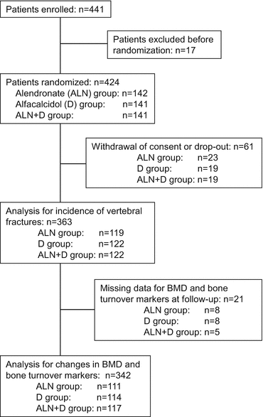
The flow chart of the disposition of the patients is shown in Fig. 7.1. Seventeen patients were excluded before randomization because fresh fractures were identified before randomization. Of the 424 randomized patients, 61 were excluded from the data analysis because of the withdrawal of consent or dropout before completion of this study. Thus, 363 patients were included in the analysis of vertebral fracture incidence. Furthermore, 21 patients were excluded from the assessment of changes in BMD and bone turnover markers because of missing data at follow-up.


Fig. 7.1
Flow chart of patient disposition
Table 7.2 shows the baseline characteristics of the study subjects. There were no significant differences among the three groups in mean age, serum NTX and BAP levels, BMD, angles of thoracic and lumbar kyphosis, number of prevalent vertebral fractures per patient, or osteophyte scores.
Table 7.2
Characteristics of the study subjects at baseline by treatment group
Variable | ALN (n = 119) | D (n = 122) | ALN + D (n = 122) | P-valuea |
|---|---|---|---|---|
Age (years) | 74.1 ± 7.0 | 75.1 ± 7.0 | 74.0 ± 7.5 | 0.449 |
Serum NTX (nmolBCE/L) | 16.6 ± 5.5 | 15.8 ± 5.5 | 16.7 ± 4.2 | 0.376 |
Serum BAP (U/L) | 36.1 ± 13.5 | 33.8 ± 12.8 | 35.5 ± 12.6 | 0.359 |
Distal radius BMD (g/cm2) | 0.276 ± 0.056 | 0.279 ± 0.058 | 0.270 ± 0.053 | 0.474 |
Angle of thoracic kyphosis (°) | 38.6 ± 10.4 | 38.6 ± 12.0 | 37.2 ± 9.8 | 0.490 |
Angle of lumbar kyphosis (°) | −22.6 ± 14.4 | −20.4 ± 15.1 | −20.6 ± 13.3 | 0.431 |
No. of prevalent thoracic vertebral fractures | 1.0 ± 1.1 | 1.2 ± 1.1 | 1.1 ± 1.2 | 0.438 |
No. of prevalent lumbar vertebral fractures | 0.6 ± 0.9 | 0.5 ± 0.9 | 0.6 ± 1.0 | 0.688 |
No. of prevalent vertebral fractures | 1.6 ± 1.5 | 1.7 ± 1.7 | 1.7 ± 1.7 | 0.811 |
Thoracic osteophyte score | 3.1 ± 2.4 | 2.8 ± 2.5 | 3.0 ± 2.0 | 0.581 |
Lumbar osteophyte score | 3.6 ± 2.1 | 3.5 ± 2.3 | 3.6 ± 2.1 | 0.999 |
Osteophyte score | 6.6 ± 3.8 | 6.3 ± 4.2 | 6.6 ± 3.7 | 0.817 |
During the 6-month treatment period, new vertebral fractures included 11 fractures in 9 ALN group patients (7.6 %), 9 fractures in 9 D group patients (7.4 %), and 3 fractures in 3 ALN + D group patients (2.5 %). No significant difference was observed among the groups in the incidence of new vertebral fractures per patient. However, the incidence of new vertebral fractures per total vertebral body examined from T4 to L5 was significantly lower in the ALN + D group (three fractures in 1708 vertebral bodies examined, 0.18 %) than in the ALN group (11 fractures in 1666 vertebral bodies examined, 0.66 %) (p = 0.029). The incidence of new vertebral fractures per total vertebral body examined was not significantly different in the D group (nine fractures in 1708 vertebral bodies examined, 0.52 %) compared with the ALN group or the ALN + D group.
The longitudinal changes in BMD of the distal radius and serum NTX and BAP levels over the 6 months are shown in Table 7.3. No significant changes of BMD from baseline were observed after treatment in any of the groups. No significant differences in BMD were observed among the groups 6 months after treatment. Serum NTX was significantly decreased compared to baseline in the ALN group and the ALN + D group (p < 0.05) but not in the D group. Serum BAP decreased significantly in all groups after treatment (p < 0.05). Serum NTX and BAP levels at 6 months after treatment were significantly lower in the ALN group and ALN + D group than in the D group (p < 0.05).
Table 7.3
BMD and bone turnover markers at follow-up and % changes from baseline
Variable | ALN (n = 111) | D (n = 114) | ALN + D (n = 117) | P-valuea |
|---|---|---|---|---|
Distal radius BMD at follow-up (g/cm2) | 0.276 ± 0.057 | 0.280 ± 0.060 | 0.272 ± 0.054 | 0.532 |
Serum NTX at follow-up (nmolBCE/L) | 12.4 ± 3.3* | 15.0 ± 4.3b | 12.8 ± 3.7c,* | <0.001 |
Serum BAP at follow-up (U/L) | 24.1 ± 7.3* | 28.0 ± 8.0b,* | 23.1 ± 8.2c,* | <0.001 |
%Δ distal radius BMD (%) | +0.34 ± 0.38 | −0.01 ± 0.37 | +0.63 ± 0.37 | 0.470 |
%Δ serum NTX (%) | −22.3 ± 2.3 | 0.6 ± 2.3 | −21.6 ± 2.2 | <0.001 |
%Δ serum BAP (%) | −28.6 ± 2.2 | −11.5 ± 2.2 | −31.2 ± 2.1 | <0.001 |
We then focused on the factors protecting against incident vertebral fractures. Patients were divided into groups with and without incident vertebral fractures (n = 21 and 342, each), and measured variables were compared between the groups. Compared with the patients without incident vertebral fractures, patients with incident vertebral fractures were significantly older and had higher baseline serum NTX levels, lower BMD, more prevalent vertebral fractures with increased thoracic kyphosis, and lower osteophyte scores (Table 7.4). Treatment regimen, baseline serum BAP, lumbar lordosis angle, percent change of BMD, percent change of NTX, and percent change of BAP showed no significant differences between the groups. Finally, multivariate logistic regression analysis showed that age, baseline serum NTX level, number of prevalent vertebral fractures, and osteophyte scores significantly affected incident vertebral fractures (Table 7.5).
Table 7.4


Comparisons of variables with or without incident vertebral fractures (VFs)
Stay updated, free articles. Join our Telegram channel

Full access? Get Clinical Tree


