Chapter 16 Spinal Cord Injury
• Patients with a spinal cord injury (SCI) are usually managed within a specialist unit, by an interdisciplinary team.
• Patients with a SCI may present at a local hospital, e.g. most new cases present to the accident and emergency (A&E) department of a district general hospital, whilst patients with an established injury may be admitted to a local hospital during a period of acute deterioration or require ongoing input following discharge from a specialist unit.
• Physiotherapy plays an essential part in the management of patients from the acute phase through to end stage rehabilitation.
• Interventions include the teaching of physical skills and coping techniques that someone with a SCI will need, to regain their independence within the community.
• Many of the techniques employed are not specific to SCI rehabilitation, but are methods widely used within cardiorespiratory, musculoskeletal, orthopaedics and neurology rehabilitation, albeit applied to the SCI population.
• Thorough assessment and re-assessment is the key to developing an appropriate and effective treatment plan.
• It must be remembered, however, that along with disturbance of motor function and sensation, many other body systems may be affected and careful consideration of the impact of therapy upon these systems is required.
Aims of rehabilitation
• A spinal cord injury is considered to be one of the most devastating conditions that can occur following a trauma.
• In seconds an individual is catapulted from a familiar life as an able-bodied person into a previously unknown situation and an environment of, in most cases, permanent disability.
• Rehabilitation therefore needs to address not only the physical aspects, but also the psychological aspects that occur following a spinal cord injury.
• Whilst in the acute stage the individual will for various reasons often not fully understand the enormity of the changes to their life.
• During mobilisation, rehabilitation and community re-integration the impact of loss of muscle power, sensation, movement, bladder, bowel, temperature control and sexual function in daily life will become clear.
• Adjustment to the injury is a lifelong process in which constant changes occur in the patient, their circumstances and needs; support thus needs to begin soon after injury (Bromley 2006).
• Rehabilitation aims to provide the individual with the knowledge, physical skills and coping strategies that are required, whilst developing their ability to regain the control of their life that may have been lost after the initial injury.
Goal planning
• Goal planning aims to place the patient at the centre of their rehabilitation programme, whilst increasing their engagement in rehabilitation activities (Kennedy et al 1991).
• It is based on the patient being an active participant in their rehabilitation and aims to recognise and utilise the patient’s strengths in order to meet their own identified needs.
• Goals are set with the rehabilitation team, specific measurable and realistic targets being defined to be achieved in an agreed time.
• Regular review and monitoring of success can become an empowering process.
• Areas of unmet needs can be recognised and addressed and the roles and contribution of the various health care professionals can be clarified.
• The need to address all areas affected by the SCI throughout rehabilitation is essential if the process is to be considered as one of learning and development of new skills and knowledge.
Functional goals of rehabilitation
• A knowledge of the anticipated functional outcome and physical independence in activities of daily living allows targets to be set to assist the patient achieve their physical goals.
• It also provides a structure for the team to work towards in conjunction with each patient and supports the goal planning framework.
• Goals are strongly influenced by the level of the spinal cord injury and the remaining innervated muscles (Appendix 16.3, number 6, p. 397).
Factors that may affect rehabilitation
Acute physical/medical management
Road side/initial management of acute trauma
• The highest proportions of patients with a SCI will have been involved in a fall or road traffic accident.
• At the scene of the injury, initial attention is focussed on maintaining the airway, breathing and circulation (ABC).
• In an actual or suspected SCI, a jaw-thrust/chin lift technique is used to maintain a patent’s airway rather than extension of the neck (ACS 2006).
• Once considered safe, extrication and transfer to the closest accident and emergency (A&E) department is conducted with ‘full body spinal protection’ established.
• This utilises a cervical collar, spinal board and head restraint (Harrison 2007).
• All unconscious or multitrauma patients with the potential for SCI are treated prophylactically as if they have injured their cord.
• The management of the individual will commence immediately to address the multisystem impairments that are secondary to SCI and to prevent avoidable complications.
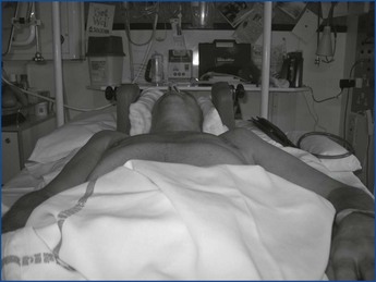
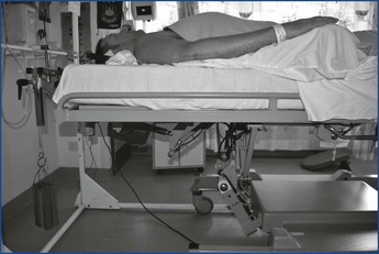
• Conservative management is followed initially, using postural reduction with or without traction to align the vertebral column.
• Respiratory support is provided if required, regular turns are instigated for the management of the skin and a regimen of treatment for the care of the paralysed bladder and bowel is commenced.
• Patients may be sedated or require admission to the intensive therapy unit (ITU) for multisystem management.
• A variety of medications may be used to control the effects of a damaged spinal cord, including those to alleviate pain, treat infections or anticoagulation therapy to prevent the formation of a deep vein thrombosis.
• It is no longer standard practice to offer high-dose methylprednisolone for spinal cord swelling (Short et al 2000), but this may still be seen in some district general hospitals.
Management of the spine
• The duration of bed rest depends on the type and cause of injury, the degree of spinal instability, the method of management, i.e. conservative or surgical and medical stability.
• The principles of management of the spine are to:
• The decision to manage the injury conservatively or surgically is multifactorial and dependent upon experience and ability of the medical team.
• There appears to be no difference in outcome between surgical and conservative management, although surgery may be associated with greater complications El Masry & Jaffray (1992).
• Patients with non-traumatic injuries are usually mobilised as soon as they are medically stable.
Conservative management
• Patients with traumatic injuries may be treated with a minimum of 6 weeks bedrest. An X-ray and computerised tomography scan (CT) of the spine will decide whether further bedrest is indicated. The total bedrest period may be 10–12 weeks in an uncomplicated case.
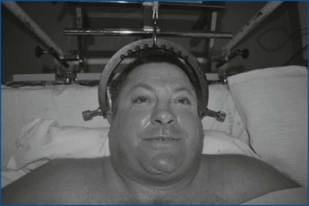
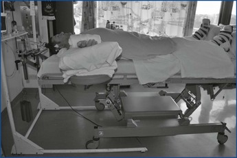
• Dislocations are usually treated with postural reduction, using pillows for the thoraco-lumbar lesions and neck roll and skull traction for the cervical lesions (Figures 16.1 and 16.2).
• Skull traction is usually maintained for 6 weeks, and is dependent thereafter on a CT taken to check the position and degree of bony callus formation.
• If further bed rest is indicated, the position of the neck will typically be maintained by using a neck roll or hard collar.
• If Halo-traction with a vest is used, the patient can be mobilised earlier, but the Halo traction will be maintained for at least 12 weeks post-injury.
Surgical intervention
• Surgical management of a traumatic injury will greatly reduce the period required on bedrest.
• The type and length of internal fixation used will vary depending on the site, number of levels involved and severity of the injury.
• The length of the fixation should be kept to a minimum by the surgeon and usually should extend just one segment above and below to prevent additional complications, e.g. reduced range of movement in the spine caused by the implant.
• The postoperative bedrest period can vary, but is typically 1 week.
Physiotherapy assessment and treatment planning
• The overall purpose of physiotherapy for patients with spinal cord injury is to improve health-related quality of life through improving their ability to participate in activities of daily life (Harvey 2008).
• The accurate identification of the factors impacting on the person’s ability to participate is achieved through assessment and re-assessment and is the key to successful management.
• Assessment tools are numerous and should be used to monitor and measure progress.
• The International Classification of Functioning, Disability and Health (ICF) can be used to facilitate the process. The reader is referred to Chapter 16 in Volume 1 (World health Assembly, 2001).
Acute physiotherapy management and rehabilitation
• The initial bed rest phase will be experienced by all individuals with a spinal cord injury.
• The duration will vary greatly dependent on cause, impairment and management.
• This period demonstrates a period where intervention is deemed essential to influence respiratory and life-threatening conditions.
• This is also an opportunity for many patients to commence rehabilitation and education, albeit in a limited capacity.
• The physiotherapeutic aims of acute management of an individual with a spinal cord injury are:
Maintenance and progression of respiratory function
• Pulmonary complications in spinal cord injury are common and are directly correlated with mortality. The higher the level of neurological injury, the more complications are likely (Bromley 2006).
• Respiratory physiotherapy aims include:
• Prophylactically patients should be offered breathing exercises, preferably using an incentive spirometer to provide ‘biofeedback’. (assisting re-education of preserved respiratory musculature)
• Mechanical ventilation is considered when the vital capacity drops below 1 litre, and is essential below 500 mL (CSCM 2005). The use of intermittent positive pressure breathing (IPPB) should be considered for preventative treatment of patients with low vital capacities. It is also a useful tool to teach the ultra high lesion how to ‘rescue breathe’ with their upper accessory muscles when off the ventilator by manipulating the ‘sensitivity’ dial.
• Abdominal binders should be used by patients without abdominal innervation when upright. This prevents the typical postural drop in vital capacity seen in the majority of SCI patients (Prigent et al 2010).
• Sputum clearance techniques should be taught to the patient or their family/carers when assistance is required. A peak cough flow (PCF) of 160 L/min is deemed essential for clearing airways, with values greater than 270 L/min ensuring a reduction in respiratory infections in the neurologically impaired (ATS 2004). This is difficult to achieve for many patients without innervation of the abdominal muscles and those with low vital capacities. When this is the case, a manual-assisted cough or use of the cough assist machine is recommended (BTS 2009).
• Respiratory deterioration can occur due to any number of reasons, e.g. ascending neurology, respiratory muscle fatigue, abdominal distension, over sedation, excessive IV infusion, respiratory infection, aspiration or even enforced smoking cessation (Dicpinigaitis et al 2006).
• When deterioration does occur, fatigue will be an issue, a ‘little and often’ approach is suggested.
• Close liaison with the medical team is necessary, the physiotherapist is encouraged to use a combination of humidified oxygen, postural drainage, breath augmentation, e.g. IPPB, manual hyperinflation or cough assist machine, and sputum clearance techniques, e.g. assisted cough timed with suction, for those whom can’t clear to the mouth.
• Early implementation of Non-Invasive Ventilation (NIV) or mechanical ventilation is advised with rising PaCO2, prior to respiratory arrest, to protect the healing spinal cord (Gardner et al 1986).
• Early tracheostomy is advocated for those difficult to wean from invasive ventilation (Harris 2007).
• Suction must be approached with caution in tetraplegia during the period of spinal shock. Unopposed vagal tone due to the parasympathetic dominance evident in this patient population predisposes the patient to bradycardia and potentially sinus arrest (RCP 2008). Pacemaker insertion should be avoided due to the fact this presentation typically resolves naturally as spinal shock passes and a pacemaker would contraindicate any future MRI scans. It is better managed with pre-oxygenation and prophylactic sympathomimetics such as glycopyrrolate. It does not contraindicate chest physiotherapy, as omission would invariably lead to respiratory failure.
• Weaning from ventilation is a team approach and should be gradual due to respiratory muscle fatigability.
• An approach of progressive ventilator free breathing has been shown to be twice as effective as the typical approach of decreasing pressure support, as used in the general population (Peterson et al 1994).
Joint management
• The outcome of the rehabilitation depends very much on maintaining adequate range of movement (ROM) and muscle length in the affected joints during the bed rest period.
• Prolonged periods of bed rest, pain, spasm, lack of regular repositioning and unopposed muscle activity can lead to muscle contracture and joint stiffness.
• The results of contracture development may include functional dependence, inhibited goal achievement, pain, pressure sores, difficulty to seat, increased carer load, increased spasms, respiratory compromise and a poor body image.
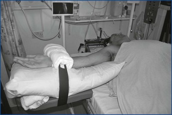
• In some cases it may be necessary to try to increase the ‘normal’ ROM in a joint to enable the patient to achieve certain functional goals later on, e.g. increased external rotation of the hip with knee and hip flexion (’tailor position’) for dressing, or increased elbow extension with wrist and shoulder extension for weight bearing without triceps innervation.
• Passive and active movements:
• Adequate analgesia needs to be provided.
• Education of the patient and family in how they can assist the process should be part of the treatment consideration.
• Close liaison with the occupational therapist is advocated to manage the special requirements of the upper limb ensuring there is co-ordination of goals and treatment.
• In order to maintain full and pain-free range in the shoulder, mobilisation of the scapula and accessory movements to the shoulder and clavicular joints are indicated prior to physiological ranging.
• While mobilising the shoulder, great care must be taken not to move the cervical spine.
• The following movements should be carried out:
• In patients with unstable lesions of T10 and below being managed conservatively or awaiting surgery, hip flexion is initially restricted to 30° in order to avoid excessive movement of the lumbar spine.
• The movement must always avoid pain at the fracture site.
• Full knee flexion is maintained with unilateral tailor position (full external rotation of the hip with limited hip flexion and full knee flexion). Rotation of the pelvis must be monitored during this movement.
• Further lower limb movements should be carried out as follows:
– Accessory movements to the metatarsal joints help to prevent deformity of the foot, which might otherwise cause pressure problems when wearing shoes and during standing.
– Adduction across midline with a straight leg.
– Abduction to the edge of the bed only (max. 45°).
– Mobilise the patella before flexing the knee.
– Extension of the hip (hip stretch) is necessary for all incomplete lesions with the potential to walk and complete lesions with the potential to use callipers, but should not be commenced without prior discussion and agreement with the consultant.
– This is achieved by placing the patient in side lying after 2–3 weeks post-injury, depending on the fracture.
– When carrying out the movement, the pelvis must be stabilised, the knee flexed to 90° with the hip in neutral rotation with no abduction or adduction.
– Particular vigilance is needed for patients with T12/L1 levels because of possible unopposed active hip flexion.
• Hamstring stretch is not usually carried out until 6 weeks post-injury because of the pull on a healing spinal cord.
• This should not be initiated without prior discussion with the consultant.
• If carried out while the patient is still on bedrest, the straight leg raise is restricted to 60°.
• There are a range of other options available to a physiotherapist to maintain joint range and function:
Maintenance of muscle power and strengthening of partially innervated muscle groups
• Post spinal cord injury a number of problems can often be identified with muscle strength within the same individual: complete paralysis, partial paralysis of a muscle and neurologically intact muscles which can be functionally weak and/or deconditioned.
• The appropriate intervention for the partially paralysed muscle or the neurologically intact muscle is the development and instigation of a strengthening programme.
• A strengthening programme can commence during the acute phase using all remaining partially innervated or intact muscles, e.g. high cervical lesions may undertake sling suspension or assisted exercises, whereas weight training/Thera-Bands may be appropriate for paraplegics.
• With all patients care must be taken to ensure excessive effort does not cause movement of the fracture site, encourage spasticity or develop unwanted compensatory strategies.
Development of desired compensatory movements

Stay updated, free articles. Join our Telegram channel

Full access? Get Clinical Tree


