SIRS, Sepsis, and Multi-system Organ Dysfunction and Failure


SIRS, Sepsis, and Multi-system Organ Dysfunction and Failure


Wendy L. Wahl


Sepsis is the body’s systemic inflammatory and coagulation response to infection. It is a life-threatening syndrome which affects tissue remote from the site of initial infection and can lead to multiple system organ failure (MSOF) and death. It is estimated that there are more than 750,000 new cases of sepsis in the United States each year with as many as 215,000 deaths annually.1 The Centers for Disease Control and Prevention ranks septicemia as the tenth leading cause of death in the United States,2 with a severe sepsis mortality estimated at 28% to 50%3 (see Fig. 1).

Before 1992, the reporting of deaths or even the incidence of sepsis was clouded by a lack of clear definitions for sepsis and associated conditions. The American College of Chest Physicians (ACCP) and the Society of Critical Care Medicine (SCCM) established standard definitions for systemic inflammatory response syndrome (SIRS) and sepsis.4 Despite this, mortality from severe sepsis is often coded as a complication of some other disease process, rather than a primary discharge diagnosis leaving the true incidence underreported.3 A sepsis consensus conference in 2001 reviewed the diagnostic criteria for sepsis to include the concept of predisposition, insult (infection), response, and organ dysfunction (PIRO).5 The four categories are summarized by the following terms:


SYSTEMIC INFLAMMATORY RESPONSE SYNDROME

SIRS is a bodily response to a wide variety of clinical insults such as tissue injury from pancreatitis, trauma, burns, blood product infusion, malignancy, endocrine disease, or infection. It is manifested by two or more of the following signs or symptoms: (a) heart rate >90 beats per minute, (b) respiratory rate >20 breaths per minute or Paco2 <32 mm Hg, (c) temperature >38°C or <36°C, and (d) a white blood cell count > 12,000 cells per mm3 or <4,000 cells per mm3 or immature (band) forms accounting for >10% of the neutrophils present. SIRS does not imply the presence of an infectious organism but is a sign of systemic inflammation.


SEPSIS

The clinical syndrome is defined by the presence of both a systemic inflammatory response and infection. In addition to the signs of SIRS, the 2001 consensus conference added mental status changes, significant edema, hypotension, acute oliguria, and tissue hypoperfusion as nonspecific signs to aid clinicians in the diagnosis of sepsis (see Table 1).


Severe Sepsis

The development of multisystem organ dysfunction (MSOD) accompanying sepsis was added for use in various sepsis assessment tools. This category includes MSOF and MSOD (to be discussed later in the chapter).


Septic Shock

The onset of acute circulatory failure with persistent arterial hypotension despite adequate fluid resuscitation
and with no other cause of hypotension characterizes septic shock. For adults, a systolic arterial pressure of <90 mm Hg or a drop of >40 mm Hg from baseline defines hypotension. For children, septic shock includes tachycardia, decreased peripheral pulses compared to central pulses, and diminished urine output.


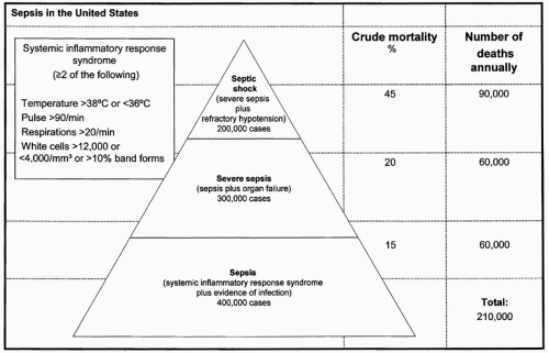
Figure 1 Sepsis in the United States. (Adapted from Manns BJ, Lee H, Doig CJ, et al. An Economic Evaluation of Activated Protein C Treatment for Severe Sepsis. N Engl J Med. 2002; 347(13):993-1000.)

Even with proper definitions in place, the diagnosis of whether or which infection is present in sepsis can still be difficult for the clinician. One or two of every three septic patients will have no growth on blood cultures or have no definite site of infection identified.3,6 Yet, the term sepsis indicates infection, and studies show that SIRS may persist even after treatment of the infectious source.7 The presence of SIRS alone does not increase the risk of death, but the deterioration of organ dysfunction seen in severe sepsis does worsen outcome.8 In a large study that followed the natural history of SIRS patients, 48% were diagnosed with an infection. Twenty-six percent of patients had uncomplicated sepsis, 18% developed severe sepsis with organ dysfunction, and 4% had septic shock. Bacteremia was more common in those with increased symptom severity, with positive blood cultures in 17% of septic patients and 69% of patients with septic shock.9


EVALUATION AND DIAGNOSIS


Physical Examination

Fever is the most common presenting symptom of sepsis and should be a signal for further clinical evaluation of infection in the patient. Elderly patients with sepsis or those with thermoregulatory dysfunction may present with hypothermia.10 Physical examination should include a rapid, global review of the patient’s condition with continuous monitoring. Patients in shock should have arterial catheters placed for blood pressure monitoring. Signs of SIRS may suggest infection or ongoing inflammation. Other clinical evidence of poor perfusion such as change in mental status, low urine output, mottling, and poor capillary refill should be evaluated in addition to blood pressure (Table 1). Sites of potential infection such as the chest, abdomen, wounds, or skin and existing catheters should be examined for obvious signs of infection. The lung, abdominal cavity, and urinary tract are the three most common sites of infection causing severe sepsis.11,12 Infections leading to sepsis can also arise in surgical sites from the skin to the deep muscle layers. Other nosocomial causes of sepsis are intravenous catheter infections, ventilator-associated pneumonia, and
sinusitis. For 20% to 30% of patients, the site of infection may not be identified.3 In patients undergoing more specialized procedures such as maxillofacial reconstruction, spine fixation or craniotomies, closed space infections of the sinuses, or intracranial space should be considered if other, more common sources are not identified as the infectious source.


TABLE 1 SIGNS AND SYMPTOMS SUGGESTIVE OF SEPSIS


Documented or suspected infection with some of the following:


General physiologic variables


Temperature >38.3°C or <36°C


Heart rate >90 beats/min or 2 SD above the mean for age


Tachypnea


Altered mental status


Significant edema or positive fluid balance (>20 mL/kg/24 hr)


In the absence of diabetes, plasma glucose >120 mg/dL


Inflammatory variables


WBC count <4,000/µL or >12,000/µL


Normal WBC with >10% immature forms


Plasma C-reactive protein >2 SD above normal value


Plasma procalcitonin >2 SD above normal value


Hemodynamic variables


Hypotension: SBP <90 mm Hg, MAP <70 mm Hg or SBP decrease of 40 mm Hg


S[V with bar above]o2 >70%


Cardiac index >3.5 L/min/M2


Organ dysfunction variables


Arterial hypoxemia: Pao2/FIO2 <300


Acute oliguria: urine output <0.5 mL/kg/hr for at least 2 hr


Creatinine increase >0.5 mg/dL


Abnormal coagulation: INR >1.5 or aPTT >60 s


Ileus (absent bowel sounds)


Thrombocytopenia: platelet count <100,000/µL


Hyperbilirubinemia: plasma total bilirubin >4 mg/dL


Tissue perfusion variables


Elevated lactate: >1 mmol/L


Decreased capillary refill or mottling


SD, standard deviation; WBC, white blood cells; SBP, systolic blood pressure; MAP, mean arterial pressure; S[V with bar above]o2, mixed venous oxygenation; PaO2, partial pressure of arterial oxygen; FIO2, fraction of inspired oxygen; INR, international normalized ratio.


Laboratory Data

Laboratory studies should include a complete blood count with differential, chemistry profile, arterial blood gas with lactate level, prothrombin time and partial thromboplastin time, and urinalysis.13 Monitoring lactate levels for guiding resuscitation has been shown to be helpful in septic patients for prognostication.14,15 Clearance of lactic acidosis with fluid resuscitation is associated with improved outcomes.

A major response to inflammatory or infectious insults is the release of cytokines. Although not routinely monitored in the clinical setting, plasma levels of tumor necrosis factor alpha (TNF)-α, interleukin (IL)-1, IL-6, IL-8, IL-10 and their soluble receptors are elevated in both infections and noninfectious SIRS. The degree of elevation correlates with the severity of disease.12 After the release of TNF-α, IL-1, and IL-6, acute phase reactant proteins, C-reactive protein (CRP), and procalcitonin (PCT) levels rise. A 10-to 100-fold increase in CRP is common in SIRS patients. Patients with systemic infections have been found to have a rise in levels of PCT, which can be predictive of sepsis and multiorgan dysfunction. Elevated PCT levels have been shown to be a better marker of sepsis than temperature, leukocyte count, TNF-α, IL-6, or CRP levels.16,17,18,19

By definition, sepsis is a response to infection. Consideration of noninfectious etiologies for SIRS should be considered including surgery, trauma, burns, hematoma, subarachnoid hemorrhage, venous thrombosis, pancreatitis, myocardial infarction, transplant rejection, thyroid storm, acute renal insufficiency, lymphoma, tumor lysis syndrome, blood products, opiates, benzodiazepines, anesthetic-related malignant hyperpyrexia, neuroleptic malignant syndrome, and erythroderma.11,20 After a thorough physical examination and consideration of noninfectious causes of SIRS, cultures and diagnostic studies should be performed to identify causative agents.


Site of Infection

Despite the problems in making the diagnosis of infection, it is important to continually examine the patient for changes. Thirty percent of septic patients may never have a causative organism identified. In some cases, it may be difficult to differentiate colonization from infection. In others, previous use of antibiotics may affect culture results. The Surviving Sepsis Campaign guidelines recommend prompt diagnostic testing. Blood cultures are indicated in patients who appear septic with fever or hypothermia, chills, leukocytosis or neutropenia, left shift of neutrophils, suspected infection, hemodynamic instability, or new renal insufficiency. The SCCM guidelines recommend that one pair of blood cultures be obtained at the onset of symptoms and once again at 24 hours.20 Subsequent blood cultures should be based on the clinician’s suspicion for ongoing bacteremia or fungemia. Two sets of blood cultures should be drawn from peripheral sites. If this is not possible, then one set should be drawn peripherally and the other from a recently inserted central catheter after careful cleansing of the port site.

Diagnosis of pulmonary infections should be based on clinical examination, chest radiographs, and inspection of the patient’s respiratory secretions. Positive findings in conjunction with alteration in leukocyte counts and changes in oxygenation are fairly suggestive of a respiratory tract infection. If the patient is intubated, we prefer diagnosis of ventilator-associated pneumonia using bronchoalveolar
lavage (BAL) and quantitative cultures to decrease the chance of treating colonization.21,22,23 Use of “mini-BAL,” where a catheter is passed deep into the airway and blindly lavaged, has been used by some, but comparison to regular BAL is recommended to ensure reliability in one’s own practice. Also, in patients with infiltrates on one side of the chest radiograph, a mini-BAL may not accurately diagnose the infectious process because it may not sample the affected side.

For hospitalized patients with central venous catheters, the catheter exit site and tunnel should be inspected for evidence of erythema or purulence. In the face of thrombosis of the catheter, purulence, embolic phenomenon, or sepsis, the catheter should be removed and cultures from the tip sent. Blood cultures from the catheter and a peripheral site are useful to determine if the infection was primarily from the catheter or related to another site of bacteremia or whether the catheter was colonized versus infected.24

Urinary tract infections are common in hospitalized patients, and urine samples should be sent for culture, microscopy, and Gram stain in febrile patients. Presence of pyuria is easily detected with leukocyte esterase testing. For patients with an indwelling Foley catheter, specimens should be taken from the urine port and not from the collection bag.24

Recent surgery adds the risk of surgical site infections as a cause of sepsis. Consideration of both superficial infection of the skin and soft tissue, as well as of the deeper layers of muscle and fascia should be made. For intracavitary infection such as intra-abdominal abscess or fascial infection, abdominal computed tomography (CT) may be necessary for detection.24 In cases of dean-contaminated or contaminated procedures, the organisms common to the organ involved in the surgery should be considered as the potential infectious flora. For clean surgical procedures, Staphylococcus aureus is the most common causative organism.18


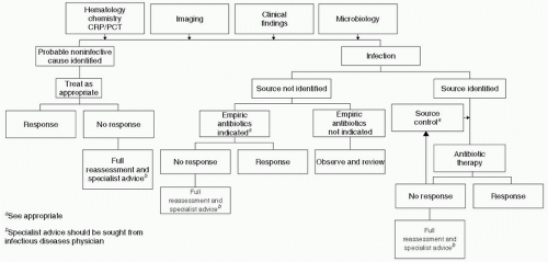
Figure 2 Algorithm for evaluation of suspected sepsis. (Adapted from Llewelyn M, Cohen J. Diagnosis of infection in sepsis. Intensive Care Med. 2001;27:S10-S32.)

Central nervous system infections should be considered in patients who have unexplained alterations in consciousness or focal neurologic signs or in recent trauma patients who have had intracranial pressure monitors or other neurosurgical interventions. A noncontrast head CT should be performed to detect contraindications to lumbar puncture (signs of high pressure). Otherwise, lumbar puncture should be performed.

For a summary of the evaluation for sepsis see Fig. 2.


TREATMENT


Antibiotics

Identification of the site of infection is a major determinant in antimicrobial therapy. Pneumonia, urinary tract infection, and bloodstream infections comprise 75% of medical nosocomial infections, and of these, 90% are related to indwelling tubes such as endotracheal tubes, Foley catheters, and central venous lines.11 These devices should be removed as soon as possible to decrease the risk of infection. However in the face of sepsis, the clinician must make the best educated guess as to the source of infection and likely pathogens. Delays in administering antibiotic therapy in septic patients have been associated with
increased mortality.25 In addition, inappropriate antibiotic therapy has also been shown to increase morbidity and mortality from survival rates of 63% to 92% for correct antibiotic therapy down to 10% to 50% for ineffective antibiotic regimens.26,27,28 When selecting empiric antibiotic therapy, clinicians should take into account the following factors: likely site of infection, likely organisms, history of recent antibiotic use (which may increase resistance), culture data if known, drug penetration into the suspected site of infection, dose, and frequency of dosing.28,29 Because the speed of delivery and the appropriateness of antibiotics affect patient outcomes, broad-spectrum coverage of likely pathogens should be empirically started. When culture and sensitivity data become available, the antibiotic regimen can then be adjusted. Lack of correct initial antibiotic coverage for offending pathogens is a primary risk for increased mortality. Even with the correct antibiotics in the face of septic shock, delays in antibiotic administration increase mortality. In 2,154 septic shock patients who received correct antibiotic therapy after onset of hypotension, each hour of delay was associated with a decrease in survival of 7.6%. Septic shock patients receiving antibiotics in the first hour after onset of hypotension had a survival rate of 79.9%, but only 50% of patients received effective antimicrobial therapy within the first 6 hours.25

To assist in choosing the correct antibiotic regimen, the clinician should categorize the likely pathogen into the following categories: (i) Gram positives, with or without Enterococcus and with or without methicillin-resistant Staphyloccus aureus (MRSA), (ii) Gram negatives, with or without Pseudomonas, Acinetobacter, or Enterobacter, (iii) atypical organisms, including Legionella, and (iv) anaerobic organisms, including Bacteroides. Prolonged hospitalization, recent exposure to antibiotics, immune status, and age contribute to the likelihood of having an infection with a nosocomial pathogen such as MRSA, Enterococcus species, resistant Gram negatives, Clostridium difficile, and Candida species.30 Knowledge of the likely pathogens in one’s own intensive care unit (ICU) is instrumental to choosing the correct antibiotic regimen because there is wide variability across ICUs even in the same institution.31


Fluids and Resuscitation

Hypotension associated with septic shock has many contributing factors. There may be large decreases of plasma volume from fluid loss into the interstitial space causing hypovolemia.32 There can be myocardial depression and decreased vasomotor tone. Despite the etiology, outcomes are improved with early fluid resuscitation. In patients randomized to early fluid boluses guided by either central venous or pulmonary artery catheter monitoring, Rivers et al. found a decrease in mortality when patients were aggressively resuscitated with fluid within the first 6 hours of presentation to the emergency department in septic shock.33 “Early goal-directed therapy” targeted a mean arterial pressure of 65 mm Hg. If after fluid administration the goal pressure was not obtained, vasopressors were added. Central venous oxyhemoglobin saturations (CvO2 sat) were followed in an attempt to achieve CvO2 of 70%. The mortality of the early goal-directed therapy group was 30.5% compared to the observed mortality of the standard therapy group of 46.5%.33 The type of fluid infused for resuscitation was not predetermined for the early goal-directed study but was left to clinician preference with the recommendation that a hematocrit <30 mg per dL be treated with red blood cell transfusion.

The debate over whether colloid or crystalloid is the best fluid for sepsis remains controversial. In an almost 7,000-patient, randomized study in Australia and New Zealand comparing use of 4% albumin to normal saline solution in patients admitted to the ICU, there was no difference in mortality, hospital or ICU days, onset of new organ failure, or mechanical ventilation for those receiving albumin compared to normal saline.34 For trauma patients, there was a trend toward improved survival with normal saline (p = 0.06), whereas in patients with severe sepsis there was a trend toward improved survival with albumin use (p = 0.09).34 In other meta-analyses, there have been mixed results where morbidity may be decreased but mortality remained unaffected with albumin use.35 However, other meta-analysis revealed no apparent difference in pulmonary edema, mortality, or length of stay between colloid and crystalloid resuscitation.36,37

Only gold members can continue reading. Log In or Register to continue

Stay updated, free articles. Join our Telegram channel

Oct 17, 2016 | Posted by in MANUAL THERAPIST | Comments Off on SIRS, Sepsis, and Multi-system Organ Dysfunction and Failure

Full access? Get Clinical Tree

Get Clinical Tree app for offline access