Chapter 4 The shoulder joint
Anatomy
2. It is the articulation between the head of the humerus (ball) and the glenoid fossa (socket) of the scapula.
3. It has a loose capsule surrounding the joint, allowing for the large range of movement and multiple degrees of freedom associated with this joint. There is an increased laxity in the capsule inferiorly, to allow for abduction at the joint.
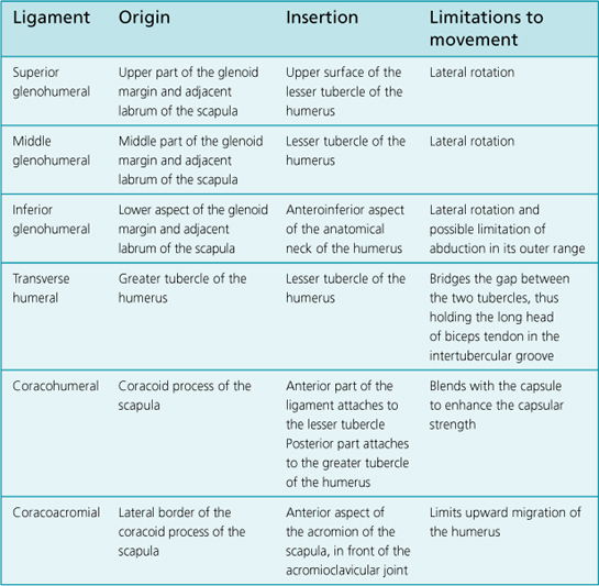
4. The joint is strengthened anteriorly by capsular ligaments (superior, middle and inferior glenohumeral ligaments).
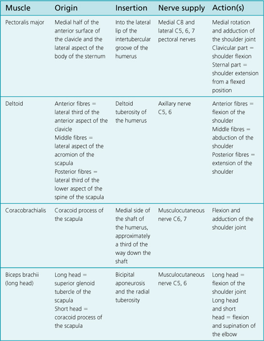
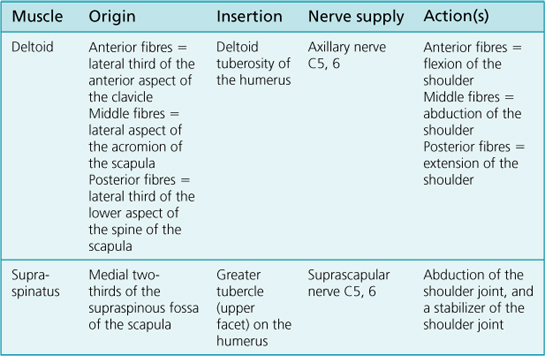
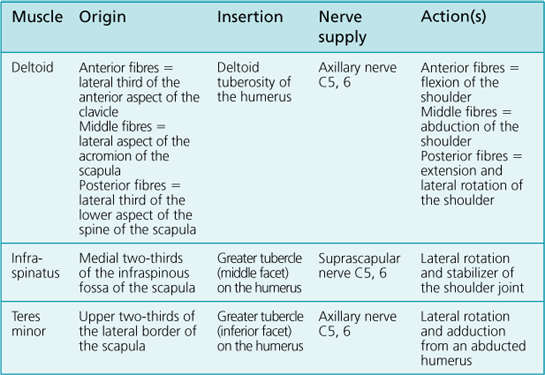
5. The joint is also strengthened by the rotator cuff muscles – supraspinatus, infraspinatus, teres minor and subscapularis.
6. The glenoid fossa is deepened by the glenoid labrum, a thick wedge of cartilage that surrounds the articular margins, increasing the congruency of the joint.
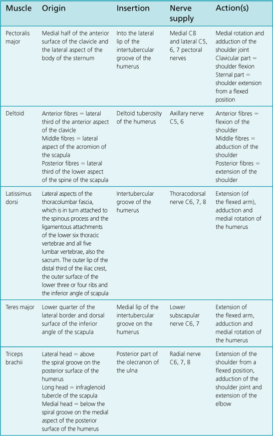
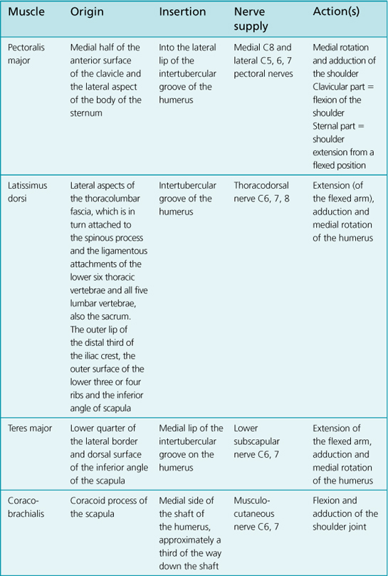
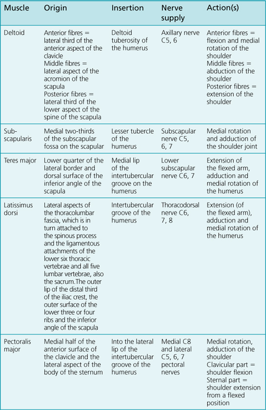
7. The movements that take place at the shoulder joint are: flexion, extension, abduction, adduction, lateral (external) rotation and medial (internal) rotation.
Bony Landmarks to be Palpated
The scapula – acromion process, acromion angle, spine of the scapula, coracoid process, inferior angle and medial border.
Measurement
Range of Movement
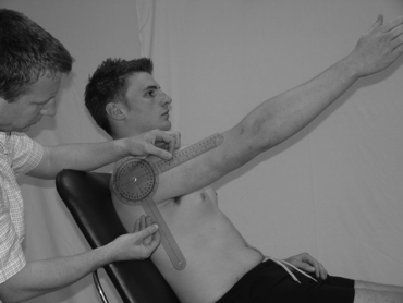
Flexion
Command to patient:
‘Keeping your elbow straight, move your arm forwards and upwards as far as you can.’
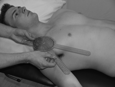
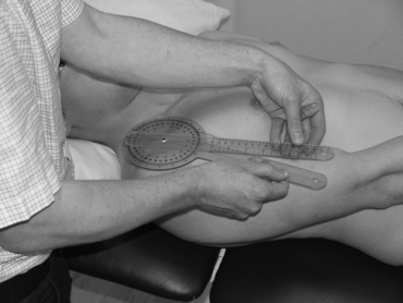
Adduction
Command to patient:
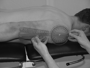
‘Move your arm across your body, as far as you can towards the opposite side of the plinth.’
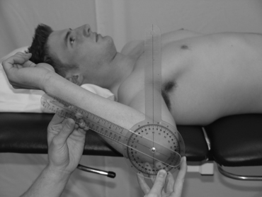
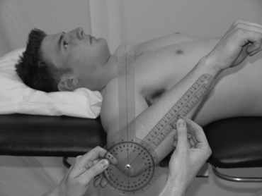
Medial (internal) rotation
Moveable arm:
This is parallel to the longitudinal axis of the ulna, pointing towards the ulnar styloid process.
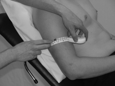
Limb Girth
Upper limb
Measurements may need to be taken of the upper limb if, for example, a patient has lymphoedema.
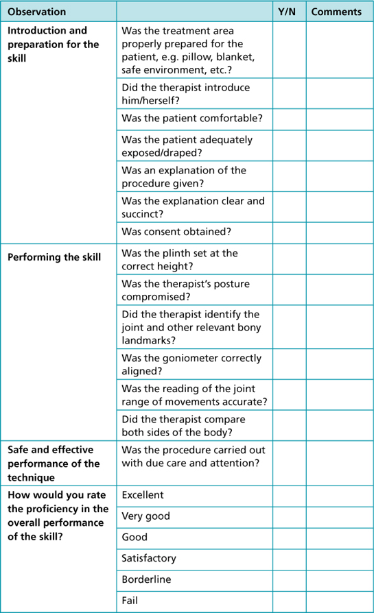
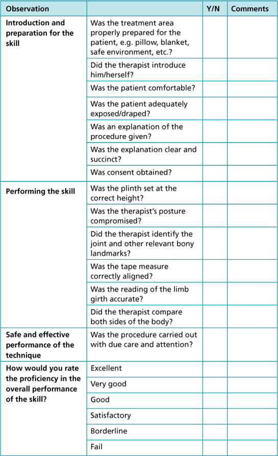
Observational/reflective checklist
Muscle Strength: Oxford Muscle Grading
Flexors

