, Maria Custódia Machado Ribeiro2 and Bruno Beber Machado3
(1)
Hospital da Criança de Brasília – Jose Alencar, Clínica Vila Rica, Brazília, Brazil
(2)
Hospital da Criança de Brasília – Jose Alencar, Hospital de Base do Distrito Federal, Brasília, Brazil
(3)
Clínica Radiológica Med Imagem, Unimed Sul Capixaba, Santa Casa de Misericordia de, Cachoeiro de Itapemirim, Brazil
Abstract
Developmental abnormalities and skeletal dysplasias comprise one of the most controversial, extensive, and complex subjects in pediatrics. This miscellaneous chapter will provide a concise review of imaging findings in selected conditions that are particularly prone to affect the immature joint. This selection is inevitably arbitrary and includes abnormalities of the bones and of the soft tissues, based mostly in their frequency, their clinical relevance, their importance for the differential diagnosis, and the potential impact of imaging for their diagnosis and management. Pseudotumoral articular and periarticular dysplastic conditions (such as focal fibrocartilaginous dysplasia and dysplasia epiphysealis hemimelica) are discussed in Chap. 6.
12.1 Introduction
Developmental abnormalities and skeletal dysplasias comprise one of the most controversial, extensive, and complex subjects in pediatrics. This miscellaneous chapter will provide a concise review of imaging findings in selected conditions that are particularly prone to affect the immature joint. This selection is inevitably arbitrary and includes abnormalities of the bones and of the soft tissues, based mostly in their frequency, their clinical relevance, their importance for the differential diagnosis, and the potential impact of imaging for their diagnosis and management. Pseudotumoral articular and periarticular dysplastic conditions (such as focal fibrocartilaginous dysplasia and dysplasia epiphysealis hemimelica) are discussed in Chap. 6.
12.2 Madelung’s Derformity
Madelung’s deformity is an uncommon anomaly characterized by shortening of the distal forearm, which presents a bayonet (or “dinner fork”) configuration, and palmar displacement of the wrist. This condition is more frequent in females in late childhood or in adolescence, presenting with pain, joint deformity, and/or limited range of motion. It may be an isolated finding or may be associated with dysplastic syndromes, like dyschondrosteosis (Léri-Weill syndrome, an autosomal dominant mesomelic dwarfism, in which Madelung’s deformity is often bilateral). Its origin is probably related to abnormal growth of the volar and medial portion of the physis of the distal radius due to the presence of an anomalous structure named Vickers’ ligament, a fibrous band that tethers the lunate and/or the triangular fibrocartilage complex to the distal radius.
Typical radiographic findings include dorsal bowing and shortening of the radius, which presents volar and ulnar angulation of its distal surface, and a triangular appearance of the distal radial epiphysis; lateral angulation and posterior dislocation of the distal ulna are also present (Fig. 12.1). The proximal carpal row has a wedge-like configuration, conforming to the altered shape of the distal surfaces of the radius and ulna (Fig. 12.1). Vickers’ ligament itself has been described with ultrasonography (US), computed tomography (CT), or magnetic resonance imaging (MRI), mostly with the latter, inserting at the gutter-like deformity of the distal radius (Fig. 12.2).
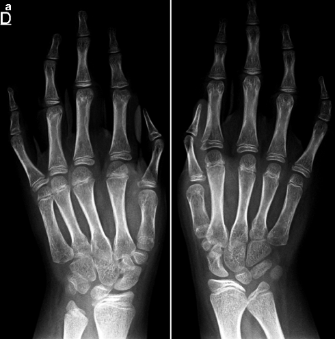
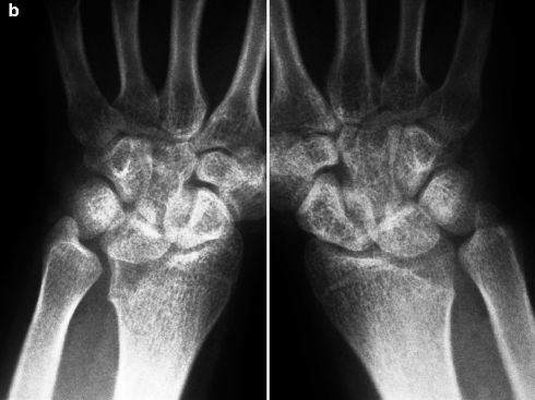
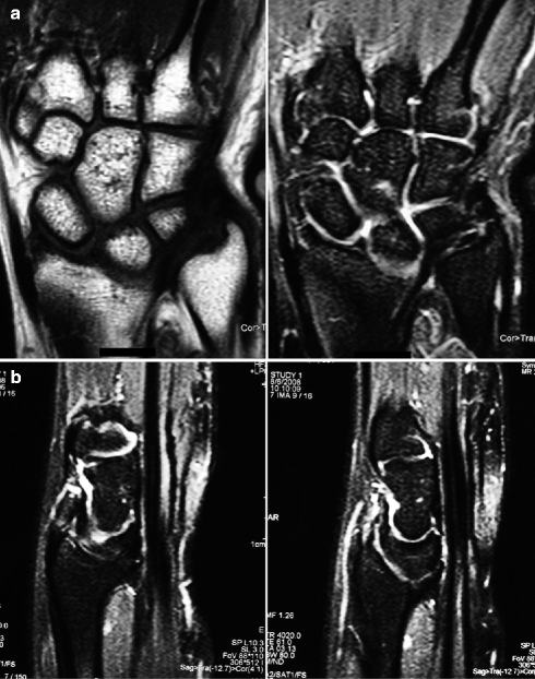


Fig. 12.1
Radiographs of hands and wrists of a child (a) and radiograph of the wrist of an adolescent (b), both with bilateral Madelung’s deformity. These images show medial inclination of the distal articular surfaces of the radii, with volar deviation more prominent in (b). The typical ulnar deformity is more marked in (a). There is abnormal alignment of the bones of the proximal carpal rows, allowing them to conform to the altered configuration of the distal epiphyses of the forearms. The distal radial epiphyses appear triangular in (b)

Fig. 12.2
Coronal T1-WI and fat sat T2-WI (a) and sagittal fat sat T2-WI (b) of the left wrist of a patient with Madelung’s deformity. The typical appearance of the distal radius is quite evident, with medial and volar angulation of the articular surface and bayonet-like configuration. Vickers’ ligament appears as a band of low signal intensity in both sequences tethering the triangular fibrocartilage complex and the lunate bone to the deformed radial epiphysis. A skin marker can be seen adjacent to the ulnar styloid, indicating a palpable lump due to dorsal subluxation of the distal ulna
12.3 Proximal Femoral Focal Deficiency
The term proximal femoral focal deficiency (PFFD) refers to a rare congenital anomaly that classically presents abnormal development and shortening of the proximal femur and deficient development of the hip joint, occurring unilaterally in most patients. PFFD encompasses a broad spectrum of developmental changes, varying from hypoplasia to complete aplasia of the proximal femur; the severity of the acetabular dysplasia correlates strongly with the extent of the deformity of the femoral head, which is often associated with subtrochanteric varus deformity. Other malformations of the affected limb may also be found, such as tibial shortening and fibular hemimelia.
Until the advent of modern imaging methods, diagnosis and staging were based on radiographic findings and on conventional arthrograms. Aitken’s radiographic classification subdivided PFFD in subtypes, varying from A (less severe, characterized by a short femur with a well-formed femoral head, normally articulated with the acetabulum) to D (very severe, with absence of the femoral head and of the acetabulum) (Fig. 12.3). However, radiographs are unable to demonstrate the non-mineralized epiphyseal cartilage and the non-ossified connection that may be present between the femoral head and the distal portion of the bone (Figs. 12.3 and 12.4). MRI is the most important imaging method for preoperative assessment of PFFD, mostly in small children, as it is able to demonstrate noninvasively the anatomy of the abnormal femur, rendering conventional arthrograms obsolete. Furthermore, MRI can discriminate if the connection between the diaphysis and the femoral head is osseous (signal intensity similar to that of the normal bone), fibrous (hypointense in all sequences), or fibrocartilaginous (hyperintense on T2-WI, similar to the cartilage). The integrity of the physis is also accurately assessed: absence of the provisional calcification zone has been described as an indicator of physeal dysfunction (Fig. 12.5). In addition, MRI can assess the articular relationships and demonstrate other osseous and soft-tissue abnormalities. Even though US is also potentially useful, MRI provides a global assessment of the joint (and of the limb) that US is unable to offer, especially considering the complex and multiple malformations often associated.
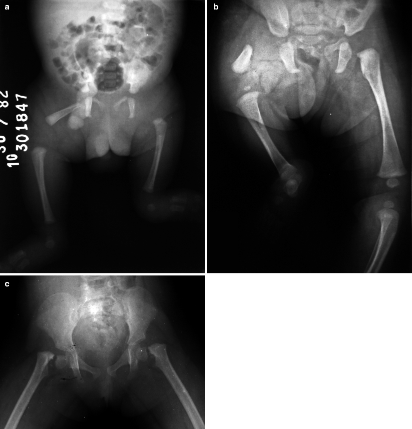
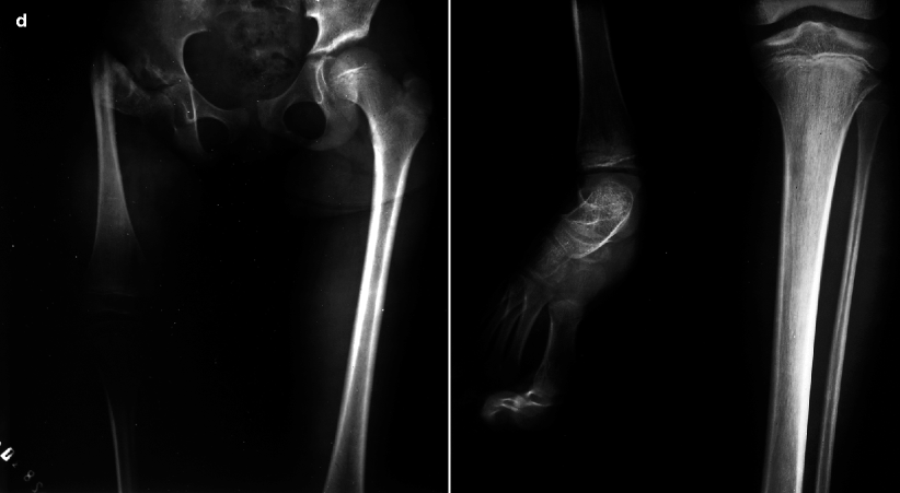
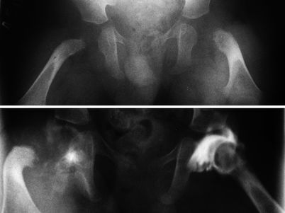
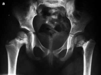
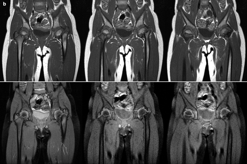


Fig. 12.3
Radiographs of the lower limbs of different patients with PFFD demonstrating the variable severity that this condition may present. In (a), both acetabula are markedly dysplastic, with no signs of ossification of the left femur and homolateral thigh shortening; the ipsilateral fibula is also malformed. In (a and b), the right femora are short and present tapering, pointed upper ends, notably in the latter image, which also displays homolateral acetabular dysplasia, cranial migration of the femoral diaphysis, and absence of the right fibula. In (c), there is symmetric and bilateral varus deformity of both femora, with mildly shallow acetabula and cone-shaped femoral heads. Figure (d) shows shortening and subtrochanteric varus deformity of the right femur, with acetabular dysplasia and reduced size of the homolateral femoral head; the right fibula is absent

Fig. 12.4
In the upper image, pelvic radiograph reveals shortening and bending of both femora, with bilateral metaphyseal irregularity. The acetabula are reasonably well formed, an indirect sign of the presence of the femoral heads. However, as the epiphyseal cartilages are not yet ossified, this study is unable to provide accurate information about them. Conventional arthrograms (lower image) delineate the contour of the femoral epiphyses, confirming that they are really present and contained within the acetabula. These images have only historical value, as arthrograms became obsolete after the advent of MRI


Fig. 12.5
Pelvic radiograph (a), coronal T1-WI (b, upper row), and post-gadolinium fat sat T1-WI (b, lower row) of the hips of a child with PFFD. Radiograph shows deficient development of the femoral head and neck at left, with homolateral varus deformity; at right, there is sclerosis and irregularity of the proximal femoral metaphysis. MRI demonstrates that the provisional calcification zone is present at right, seen as a metaphyseal line of low signal intensity, and ill-defined at left
12.4 Developmental Dysplasia of the Hip
Instead of a single disease, developmental dysplasia of the hip (DDH) comprises a wide spectrum of congenital malformations that have in common an abnormal relationship between the femoral head and the acetabulum. These conditions may vary from stable and mildly dysplastic hips to frankly dysmorphic and dislocated coxofemoral joints. Females are affected in the overwhelming majority of the cases, and bilateral disease occurs in approximately 20 % of the patients. The diagnosis is essentially a clinical one, which is usually suspected immediately after birth; delayed recognition is associated with worse prognosis and long-term sequelae (Fig. 12.6). Nevertheless, most “dislocatable” hips (especially those detected in the first 4–6 weeks of extrauterine life and not associated with an abnormally shaped acetabulum) are deprived of significance and will stabilize spontaneously, resulting from physiologic postnatal tissue laxity. There is no evidence to support disseminated use of imaging in the screening of DDH, as there is a wide “gray zone” of findings of uncertain significance between the normal hip and clear-cut DDH, so that excessive use of imaging may increase the rate of false-positive diagnoses. Indications for imaging evaluation include, among others, inconclusive physical examination, preoperative assessment, and follow-up of treatment.
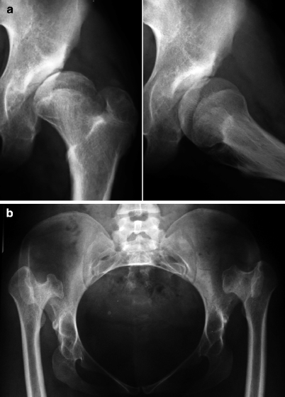

Fig. 12.6
In (a), radiographs of the left hip of a child with DDH shows articular incongruity, with a shallow and dysplastic acetabulum associated with a dysmorphic and laterally subluxated femoral head. In (b), pelvic radiograph of a 28-year-old female with bilateral DDH shows bilateral coxa vara and posterosuperior dislocation of the femoral heads, which are markedly deformed. Both acetabula are poorly developed
The usefulness of radiographs for early diagnosis is quite restricted; nevertheless, markedly dysmorphic acetabula and frank dislocations may be evident even in newborns (Fig. 12.7). Considering that the femoral heads begin to ossify only after 4–6 months of extrauterine life, radiographs are very limited in small children, as the epiphyseal cartilages will be undetectable until then. There are some classic lines drawn in the anteroposterior view of the pelvis that are used to estimate the relationships of the hip joint. Shenton’s line is an imaginary arch connecting the medial contour of the femoral neck with the superior contour of the obturator foramen; discontinuity of this arch is an indicator of joint dislocation. Hilgenreiner’s line is horizontal and drawn tangentially to the superior aspect of both triradiate cartilages, while Perkins’ line is perpendicular to the former, tangential to the lateral edge of the acetabulum. In normal children, the femoral heads (or their expected location) should be inferior and medial to the intersection of Hilgenreiner’s and Perkins’ lines, and lateral/superior migration is indicative of dislocation (Figs. 12.7 and 12.8). In dysplastic hips, the femoral head may be smaller and/or show delayed ossification if compared to the normal contralateral one (Fig. 12.8).
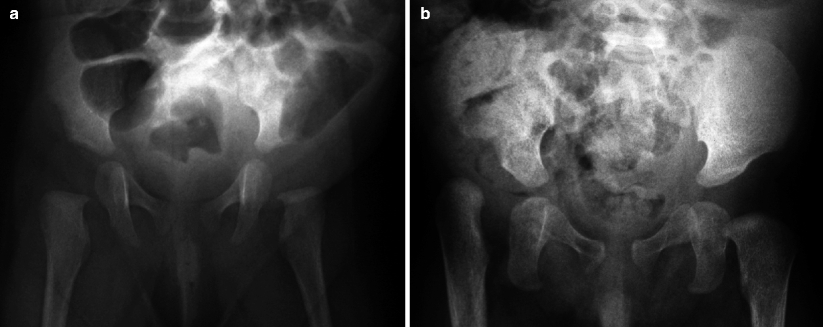
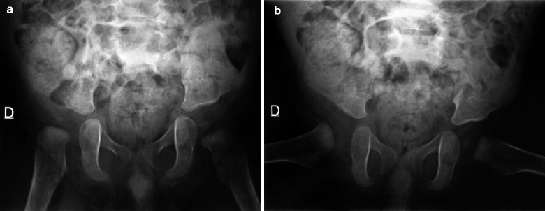

Fig. 12.7
Pelvic radiographs of two distinct patients with DDH. Shenton’s line is broken in both hips in (a), with mild lateralization of the right metaphysis and homolateral acetabular hypoplasia. In (b), the right hip shows signs of frank dislocation, with cephalic migration of the femur and hypoplasia of the acetabulum; discontinuity of Shenton’s line is seen in the left hip, as well as mild acetabular hypoplasia. The real position of the femoral heads can be only inferred at radiographs, as they are not yet ossified

Fig. 12.8
DDH at right. Pelvic radiographs reveal that the ossified portion of the right femoral epiphysis is smaller and presents delayed development if compared to the contralateral one. The right epiphysis is out of the inferomedial quadrant delimited by the intersection of Perkins’ and Hilgenreiner’s lines. Shenton’s line is clearly discontinuous at right, with hypoplasia of the homolateral acetabulum
US is the preferred imaging method for diagnosis and follow-up of DDH during the first 6 months of life, becoming gradually less sensitive and more difficult to perform as the femoral head ossifies. This imaging modality is largely used to confirm reduction of dislocation in children treated with orthotic devices, such as the Pavlik harness, being also able to evaluate joint stability in real time. However, factors such as subjective interpretation, limited reproducibility, and operator-dependent nature must be kept in mind, as well as the relatively high rate of false-positive diagnoses. The static Graf method is widely disseminated, using two angles drawn in a single coronal image for assessment of DDH (Fig. 12.9), emphasizing the morphology of the hip. The alpha angle measures the osseous acetabular roof angle (slope of the acetabulum), while the beta angle, less important, is indicative of the acetabular cartilaginous roof coverage. There is mild dysplasia if the alpha angle is between 50 and 59°, moderate between 43 and 50°, and severe if the angle has less than 43° (Fig. 12.9). The normal beta angle has less than 55°. The original classification proposed by Graf is complex, with several subdivisions based on the appearance of the anatomic landmarks and on the values of alpha and beta angles. Nevertheless, recent works have recommended a simplified approach, emphasizing descriptive terms related to the dysplastic changes and to the degree of dislocation of the femoral head, with less emphasis in the angles, given their limited reproducibility. The dynamic (real-time) technique proposed by Harcke is popular in the USA, allowing for assessment of hip stability by examining the joint in rest and under stress (pushing the femur posteriorly) in both the coronal and transverse planes; in normal patients, only minimal posterior subluxation of the femoral head is acceptable under stress.
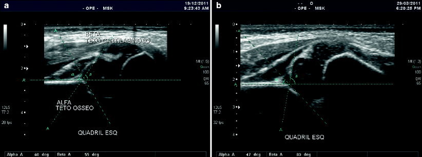

Fig. 12.9
In (a), US of the left hip of a newborn reveals normal values for the angles measured with Graf’s static method (see text). In (b), DDH is present, with reduction of the alpha angle and increase of the beta angle
CT and MRI are exceptionally used in the assessment of DDH. The most important indications include presurgical planning and follow-up of patients with casts after surgery or closed reduction to confirm that the procedure was successful (Fig. 12.10). MRI is also able to demonstrate hypertrophy and/or interposition of soft-tissue structures that may occasionally preclude closed reduction. Avascular necrosis is a potential complication of reduction, and MRI is the optimal imaging method for its diagnosis.
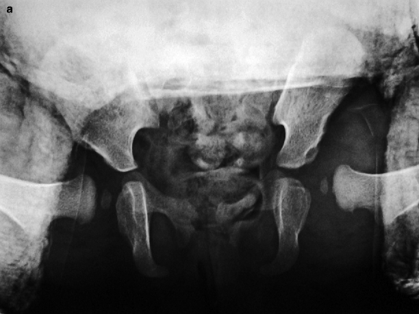
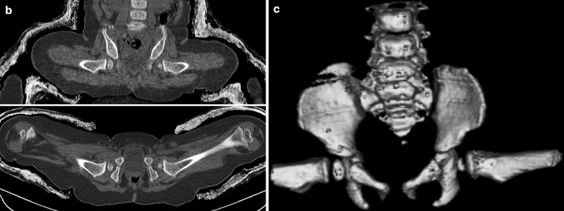


Fig. 12.10
Even though marked acetabular hypoplasia and asymmetric growth of the femoral heads are clearly seen, radiographs alone are unable to provide an accurate evaluation about how successful was closed reduction of hip dislocation after cast placement (a). CT of a distinct patient who underwent a similar procedure displays the articular relations in a safe and noninvasive way, in multiplanar reformatted images (b) and volume-rendered reconstruction (c); left-sided hypoplasia of the acetabulum and of the femoral head are present, more evident in (c)
12.5 Dorsal Defect of the Patella
The dorsal defect of the patella (DDP) is an anatomic variant that is present in approximately 1 % of the population. Although often asymptomatic, DDP may occasionally cause pain and patellofemoral symptoms. The defect is typically superolateral, along the articular surface of the bone, appearing on radiographs and CT as a small, well-delimited cortical lucency that is often surrounded by sclerosis (Figs. 12.11 and 12.12). MRI discloses a focal depression of the osseous surface that may be occasionally surrounded by bone marrow edema. The overlying cartilage is most often intact, although in some cases thinning or fraying may be found (Fig. 12.12).
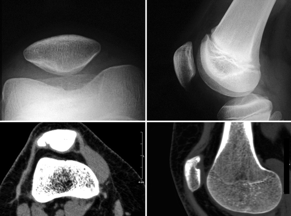
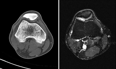

Fig. 12.11
Radiographs (upper row) and CT images (lower row) of two distinct patients with DDP demonstrates similar findings: a well-defined cortical defect in the superolateral portion of the articular surface of the patella, with subtle depression of the overlying cartilage on CT (Courtesy of Dr. Pablo Picasso Coimbra, Clinica Boghos Boyadjian, Fortaleza, Brazil)

Fig. 12.12
Transverse CT image (left) and fat sat T2-WI (right) of the right knee of the same patient. There is bone sclerosis adjacent to the superolateral cortical defect, more evident on CT, as well as thinning of the articular cartilage, whose signal intensity is heterogeneous on MRI. Dorsal defect of the patella
12.6 Transient Lateral Patellar Dislocation
The term transient lateral patellar dislocation (TLPD) refers to an acute luxation of the patella, which presents spontaneous and immediate reduction in most cases and frequently goes unnoticed. It is more frequent in children between 9 and 15 years of age. Anatomic variants – such as patella alta, lateral patellar subluxation, lateral patellar tilt, and shallow/dysplastic trochlea – are frequently found, predisposing to chronic patellofemoral instability and recurrent dislocations.
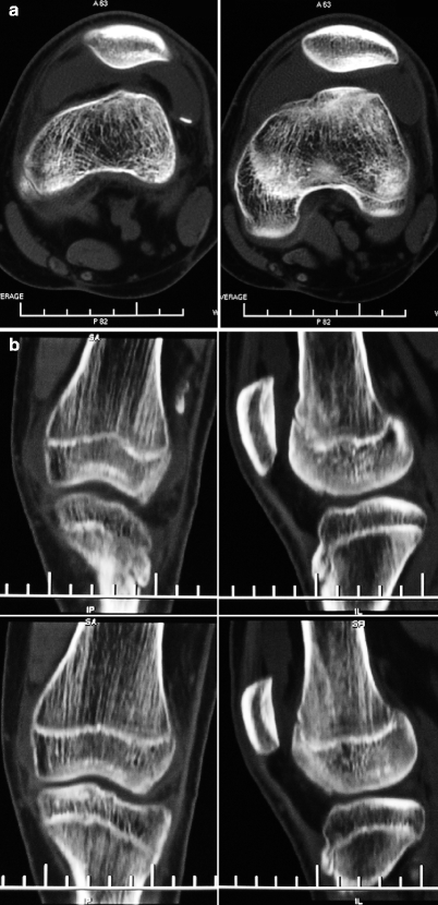
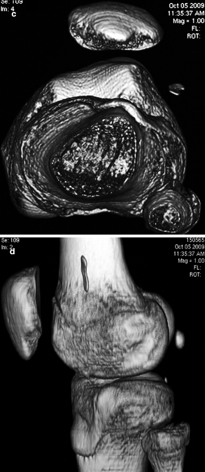
Radiographic findings are subtle and include synovial effusion and soft-tissue swelling, which are often associated with abnormal patellofemoral morphology; avulsion fractures and/or cortical deformities may also be present, better demonstrated with CT (Figs. 1.15 c, d, 12.13, and 12.14). MRI is the best imaging modality for assessment of TLPD: some classic signs allow to diagnose previous dislocation even if the articular relationships are normal when the study is performed. Joint effusion is very common, often hemorrhagic. Areas of bone marrow edema at the inferomedial portion of the patella and along the external surface of the lateral femoral condyle (“kissing contusions”), chondral/osteochondral lesions, injury of the medial soft-tissue restraints of the patella, and dysplastic changes of the patellofemoral joint are indicative of TLPD, especially when found in conjunction (Figs. 12.15 and 12.16). Bone marrow edema pattern may still be present several months after the episode of instability, even if there is complete involution of soft-tissue edema (Fig. 12.16). Intra-articular loose bodies are often present (Figs. 1.15, 12.13, and 12.15) and injuries of ligaments and menisci may be found in almost one-fourth of pediatric patients. The aforementioned predisposing structural abnormalities of the patellofemoral joint are better seen with CT and MRI (Figs. 1.15, 12.13, 12.14, 12.16, and 12.17). Many angles, lines, and relations have been described for assessment of patellofemoral instability in adults, but for most of them, there is insufficient evidence to support their use in pediatric patients.

