Scaphotrapeziotrapezoid Joint Fusion

48 Scaphotrapeziotrapezoid Joint Fusion


Indications


Long-standing scaphotrapeziotrapezoid (STT) arthritis recalcitrant to nonoperative treatment


Technique


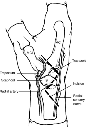
  • Dorsoradial incision that begins at Lister’s tubercle, traverses the anatomical snuffbox, and ends at the STT joint. Incision can be curved or designed with a zigzag configuration. Exposure can also be accomplished via a dorsal transverse incision (Fig. 48-1).
  • Protect the radial sensory nerves and lateral antebrachial cutaneous branches using loupe magnification. The dorsal approach exposes the STT joint between the extensor carpi radialis longus and brevis tendons through a transverse capsular incision. The radial approach is more extensive and performed between the extensor pollicis brevis (EPB) (first compartment) and extensor pollicis longus (EPL) (third compartment) tendons (Fig. 48-2).
  • The deep branch of the radial artery resides just proximal to the STT joint and must be protected in both approaches.
  • Using the radial approach, a longitudinal arthrotomy is performed from the radial styloid to the distal scaphoid (S). The distal scaphoid, trapezium (Tm), and trapezoid (Td) are identified. The articular and subchondral surfaces of the scaphoid, trapezium, and trapezoid are removed until cancellous bone is exposed (Fig. 48-3).
  • A rongeur and small curved osteotomes are employed to prevent thermal necrosis and maximized bone carpentry. Certain patients will have extremely hard bone, and a high-speed bur will be required.
  • Two to three double-ended trocar-tipped 0.45 in. (1.1 mm) wires are preset into position after the bony surface has been prepared and prior to bone grafting. This can be accomplished using one of two techniques; the wires can be driven through the trapezium or trapezoid from within the STT joint into a percutaneous position or the wires can be placed from a percutaneous position through the trapezium or trapezoid (Fig. 48-4).
  • The wire positions are adjusted to ensure passage across the arthrodesis site and engagement of the scaphoid. The wire lengths are adjusted to reside just within the trapezium or trapezoid bone. Accurate placement of the wires is essential because subsequent bone grafting will obscure visualization of wire alignment.
  • Bone graft is harvested from the distal radius from either a second transverse incision over the distal radius or a proximal extension of the radial zigzag incision.
  • The bone graft is packed into the depths of the fusion site to maintain the external dimensions of the STT joint. The preset wires are advanced into the scaphoid to secure the arthrodesis site (Fig. 48-5).
  • Additional bone graft is densely packed into the spaces between the scaphoid, trapezium, and trapezoid. The position of the wires is verified using fluoroscopy from AP, lateral, and oblique projections.
  • The pins are cut beneath the skin.
  • A radial styloidectomy is performed to prevent radial styloid impingement. The styloid is exposed by subperiosteal elevation of the first compartment. Removal of 5 to 8 mm is performed to relieve scaphoid impingement without detachment of the radioscapho-capitate or radiolunate ligament.

image

Only gold members can continue reading. Log In or Register to continue

Stay updated, free articles. Join our Telegram channel

Mar 21, 2017 | Posted by in MUSCULOSKELETAL MEDICINE | Comments Off on Scaphotrapeziotrapezoid Joint Fusion

Full access? Get Clinical Tree

Get Clinical Tree app for offline access