46 Scaphoid Excision with Capitolunate Triquetrohamate Arthrodesis
Indications
- Radioscaphoid and midcarpal arthritis with preservation of the radiolunate joint. This pattern is typically seen in long-standing scapholunate dissociation or scaphoid nonunion.
- Some surgeons prefer scaphoid excision with capitolunate triquetrohamate (CLTH) arthrodesis to proximal row carpectomy in patients with radioscaphoid arthritis and a midcarpal joint that has no evidence of degenerative changes.
Pitfall
Elderly patients and those with severe wrist stiffness have less predictable pain relief and recovery of motion.
Technique
- A dorsal, longitudinal 6 to 8 cm skin incision is made centered over the capitate (C).
- Dissect to the wrist capsule between the second and fourth compartments distal to the extensor pollicis longus tendon.
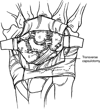
Figure 46-1
Pearl
Incise the distal edge of the extensor retinaculum between the third and fourth compartments to the level of Lister’s tubercle to facilitate exposure.
- Make a transverse capsulotomy across the entire dorsal wrist at the level of the capitolunate joint and reflect the capsule both proximally and distally from the carpus (Fig. 46-1).
- Remove the scaphoid (S) piecemeal with a rongeur.
- Excise distally projecting osteophytes from the radial styloid.
Pitfall
Avoid injury to the radioscaphocapitate ligament, which passes over the palmar surface of the scaphoid waist.
- Complete the dorsal exposure of the capitate, hamate (H), lunate (L), and triquetrum (T).
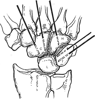
- Remove the cartilage and subchondral bone from the intercarpal articulations of the hamate, capitate, triquetrum, and lunate, with the exception of removing only the dorsal half of the capitohamate joint (Fig. 46-2).
- Prepare bone graft from the distal radius or from the iliac crest.
- Insert 0.045 in. K-wires in the carpal bones (two in capitate, two in triquetrum, and one in hamate), checking position via fluoroscopy (Fig. 46-2).


