Scaphocapitate Fusion with Lunate Excision

58 Scaphocapitate Fusion with Lunate Excision


Indications


Kienböck disease with the following:


  1. Stage IIIB disease. (See Table 58-1.)
  2. Stage IIIA disease with neutral or positive ulnar variance
  3. After unsuccessful joint leveling procedure

Technique


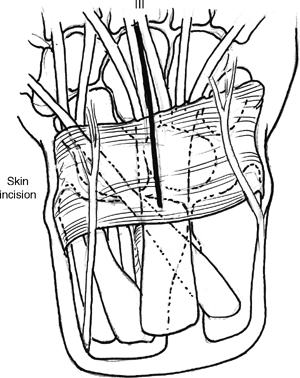
  • Longitudinal dorsal incision that begins just ulnar to Lister’s tubercle and extends to the base of the third metacarpal (Fig. 58-1).
  • Protect the radial sensory nerves and lateral antebrachial cutaneous branches using loupe magnification. Elevate flaps at the level of the extensor retinaculum.
  • The extensor pollicis longus (EPL) tendon is identified and the third compartment opened. The EPL tendon is transposed in a radial direction from the third compartment.
  • The second compartment is elevated in a radial direction and the fourth compartment in an ulnar direction. The posterior interosseous nerve is located under the fourth compartment and resected.
  • A capsulotomy is performed to expose the underlying radiocarpal and midcarpal joints. The capsular incision is created along the dorsal radiocarpal and intercarpal ligaments. This capsulotomy incision provides adequate carpal exposure (Fig. 58-2).
  • Synovectomy is performed to facilitate visualization. The lunate (L) is examined for fragmentation and collapse. A grossly deformed lunate is removed using a rongeur; otherwise, the lunate can be retained.
  • The articular and subchondral surfaces between the scaphoid (S) and capitate (C) are removed using curettes, rongeurs, and thin osteotomes. The volar rim of articular surface is not violated to maintain midcarpal dimensions (Fig. 58-3).
  • Bone graft is harvested from the distal radius. Iliac crest bone graft is an alternative option.
  • Prior to fixation, any intercarpal malalignment should be corrected. The scaphoid should align ~45 degrees relative to the radius in the sagittal plane. This reduction can be accomplished by manual pressure or by using 0.45 to 0.62 in. (1.1–1.5 mm) wires as joysticks.
  • Two guide wires for cannulated compression screw fixation are used for provisional fixation. These wires are directed from a radial to ulnar direction and may require a separate radial small incision. The wires are visualized across the scaphocapitate joint and driven into the capitate. An elongated radial styloid will require removal to facilitate guide wire placement.
  • The carpal reduction and wire position are verified by fluoroscopy.
  • Cancellous bone graft is packed into the depths of the fusion site within the denuded scaphocapitate surfaces.
  • The cannulated drill is placed over the guide wire with careful protection of the soft tissues. The cannulated headless screw is also placed over the guide wire and scaphocapitate joint (Fig. 58-4).
  • Additional bone graft is densely packed into the spaces between the scaphoid and capitate. The position of the screws is verified using fluoroscopy and anteroposterior, lateral, and oblique projections.
  • The capsule is reapproximated and closure is performed with the EPL tendon transposed into the subcutaneous tissues.

image

Only gold members can continue reading. Log In or Register to continue

Stay updated, free articles. Join our Telegram channel

Mar 21, 2017 | Posted by in MUSCULOSKELETAL MEDICINE | Comments Off on Scaphocapitate Fusion with Lunate Excision

Full access? Get Clinical Tree

Get Clinical Tree app for offline access