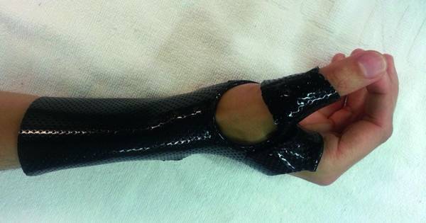
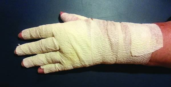
Role of Hand Therapy in the Treatment of Distal Radius Fractures Claude Poteau described fractures of the distal end of the radius with dorsal displacement of the distal fragment in 1783, but it was not until 1814 that it was published by Abraham Colles.1 Distal radius fracture (DRF) is a very common injury, accounting for almost one-sixth of all fractures occurring in developed countries.2 The various fracture patterns of the distal radius generally occur to the outstretched hand as a result of a fall. Given that the wrist is one of the most complicated joints in the human body, it is necessary to know not only its fracture patterns but also its biomechanical performance to establish appropriate hand therapy intervention. The topographic shape of the distal radius articular surface functions as a specialized support base for the carpus. To perform this function, the articular surface must be smooth, levelled, and positioned correctly relative to that of the wrist. During treatment, hand therapists should remember that there are extrinsic and extensor muscle flexors that cross the wrist and that activate strength vectors during the gripping and flexing motions of the hand. Distal radius fractures may be managed with or without surgical intervention. Both methods yield good and reliable results when used appropriately. However, in both cases, an appropriate hand therapy approach based on scientific evidence and knowledge of wrist biomechanics is needed to optimize the result. Despite the high incidence of DRF and the significant time patients with these fractures spend in hand therapy, the types and effectiveness of therapy interventions need to be widely studied to identify the practical patterns used in the conservative and postsurgical treatment. This chapter focuses on specific aspects of treatment in conservative and postsurgical approaches for restoring hand function relating to edema, pain control, restoration of motion, strength, and outcome measurements. In an acute trauma, edema fluid and hematoma surround the interosseous muscles. Capillary compression and venous stasis intensify the congestion. A tight cast further impedes venous drainage and increases the pain, meaning that the patient cannot move their fingers effectively, which may result in an intrinsic contracture. This may also result from inadequate immobilization where metacarpophalangeal joints are blocked. It is important to observe acute or postsurgical immobilization in DRFs, verifying that the patient can completely flex the metacarpophalangeal joint and that there are no compression points (▶ Fig. 27.1). Fig. 27.1 Immobilization of distal radius fracture with metacarpophalangeal joints kept free. Tendons, nerves, and vascular structures are confined and closely approximate the underlying distal radius. Edema is a part of the normal inflammatory response that occurs after trauma or surgical intervention. Edema after trauma or surgery is caused by accumulation of excessive fluid in the intercellular spaces.3 Problems occur when edema persists beyond the inflammatory phase (3–5 days). At this stage, the excess fluid or transudate consists mainly of water and dissolved electrolytes.4 Edema control in this phase should be relatively easy as long as the principles of compression, elevation, cold, and gentle active motion are observed. Compressive wraps such as Coban (3M, St. Paul, Minnesota, United States) (▶ Fig. 27.2) and elasticized sleeves or gloves can also help. Fig. 27.2 Coban compressive wrap. Therapists must be careful as some of the arm positions used for elevating the hand can create other problems such as a shoulder impingement if the patient keeps their shoulder in internal rotation and elevation for too long. Therapists should make the patient aware of this potential problem and how the limb can be correctly elevated to avoid it. Gentle active motion of the shoulder, elbow, and fingers will also help at this stage to reduce edema, but excessive exercise and heat are counterproductive.3 Cold is especially helpful during the initial inflammatory phase and performs better than contrast baths in reducing edema. The contrast bath procedure may increase superficial blood and skin temperature, but evidence of the impact on edema is conflicting and no relationship between physiological effects and functional outcome has been established. We can use different cold modalities such as cold gel pack, cryopress, or continuous ice water and cold immersion when scarring and immobilization allow it, using repeated applications every 10 minutes. Despite the benefits of cold for edema reduction, therapists must pay special attention to vascular status and in those cases where an artery has been repaired or there is a peripheral nerve involvement. Between 2 and 6 weeks there is increased capillary growth, fibroblast proliferation, and new collagen synthesis. If edema persists in this proliferative phase, it becomes more viscous and the lymphatic system can become overwhelmed by persistent edema, resulting in a sustained dynamic insufficiency. Due to an overload in the lymphatic system, the transport capacity in the lymphatic structures is reduced, and after 2 weeks this may cause protein-rich edema in the interstitium.5 This is because distal-to-proximal massage or manual edema mobilization (MEM) treatment programs in this phase help reduce edema and consequently reduce the rate of pain and increase the range of motion. Active motion, tendon gliding exercises, and orthotic and compression techniques should also be used to minimize adhesions between tissues.3
27.1 Introduction
27.2 Acute Intervention: General Considerations
27.2.1 Casting Control
27.2.2 Edema Control
Related posts:
 Undisplaced Scaphoid Fractures
 Proximal Row Fractures (Other Than Scaphoid and Pisiform Fractures): Triquetrum and Lunate Fractures
 Intra-articular Fractures of the Distal Radius (AO types C3, with Special Focus in C3.3), Open Approach
 Intra-articular Fractures of the Distal Radius (AO type C3), (Dry) Arthroscopic Approach
 Galeazzi Fracture-dislocation
 Unstable Ulnar Styloid Fracture
![]()
Stay updated, free articles. Join our Telegram channel

Full access? Get Clinical Tree


