Fig. 30.1
Schematic representation of the fracture zones in the proximal fifth metatarsal bone
30.4.1 Tuberosity Avulsion Fracture (Zone I)
Tuberosity avulsion fractures are the most common acute fracture of the proximal fifth metatarsal (Lawrence and Botte 1993).
This fracture was once thought to occur via avulsion of the tuberosity by the contracting peroneus brevis muscle during inversion of the hindfoot. However, a cadaveric study indicated that the firm attachment of the lateral band of the plantar aponeurosis is more likely than the peroneus brevis to cause tuberosity avulsion fractures (Richli and Rosenthal 1984). As yet, there is no consensus regarding the mechanism of this fracture.
Lawrence reported that fractures in Zone I have an excellent healing potential and should be treated symptomatically with a hard-soled shoe, a cast, or, in some patients, a protective wrap or brace (Lawrence and Botte 1993; Nunley 2001). Most fractures heal or become asymptomatic within 3 weeks, and radiographic union can be confirmed after 7–8 weeks (Nunley 2001; Rosenberg and Sferra 2000). Symptomatic nonunion occurs only rarely. In a prospective randomized study in which 60 patients were treated with either a short leg cast or a soft Jones dressing, all showed radiographic evidence of fracture healing by 65 days (mean, 44 days) post-treatment and returned to full weight bearing and full physical activity within 96 days (Wiener et al. 1997). However, Rettig et al. (1992) reported that eight athletes developed symptomatic nonunion of the tuberosity of the proximal fifth metatarsal. These occasional cases of symptomatic nonunion of intra-articular fracture of the tuberosity with significant articular step-off should be treated surgically (Fetzer and Wright 2006; Rammelt et al. 2004; Rosenberg and Sferra 2000; Zwitser and Breederveld 2010).
30.4.2 Jones Fracture (Zone II)
Sir Robert Jones is credited with the first description of this fracture, in 1902 (Jones 1902). The Jones fracture was defined as a transverse fracture at the diaphyseal-metaphyseal junction without extension distal to the fourth-fifth intermetatarsal articulation (Stewart 1960). Fractures in this zone take longer to heal than do more proximal fractures (Dameron 1995).
Jones fractures occur when a large adduction force is applied to the forefoot with the ankle in plantar flexion (Dameron 1995; Stewart 1960). A large bending motion produced by a high load on the plantar aspect of the fifth metatarsal head causes fracture at the junction of the proximal diaphysis and metaphysis (Fetzer and Wright 2006).
In a nonathlete who is amenable to conservative treatment, this fracture can be treated with immobilization and non-weight bearing for 6–8 weeks. A prospective randomized study found that functional treatment significantly reduced the time to return to the preinjury level relative to immobilization in a short leg cast, whereas a combination of both treatments led to complete bone healing in all patients (Wiener et al. 1997). Clapper et al. (1995) reported on a series of 235 true acute Jones fractures and found that 8 weeks of non-weight bearing followed by weight bearing as tolerated while wearing a cast was successful in 72 % of patients (mean time until union, 21.2 weeks), whereas 28 % showed clinical and radiographic evidence of nonunion 25 weeks after injury. In the seven patients in whom conservative treatment failed, intramedullary screw fixation achieved 100 % union in all patients after a mean of 12.1 weeks. True Jones fractures require a prolonged healing period when treated with 8 weeks of non-weight bearing followed by weight bearing as tolerated while wearing a cast. Dameron (1995) reported that the patient’s lifestyle and desired activity level should determine the choice of treatment of Jones fracture, which can include a functional metatarsal brace, a stiff-soled shoe, a short-leg cast, or even internal fixation with a screw.
30.4.3 Proximal Diaphyseal Stress Fracture (Zone III)
Proximal diaphyseal stress fracture involves the proximal 1.5 cm of the diaphysis. DeLee et al. (1983) defined the criteria for a stress fracture of the proximal fifth metatarsal to include (1) a history of prodromal symptoms affecting the lateral aspect of the foot, (2) radiographic evidence of a stress lesion in the bone, and (3) no history of treatment for a fracture of the fifth metatarsal.
The mechanism of this fracture usually involves stress or fatigue produced by repeated normal loading of the plantar surface of the fifth metatarsal head over a relatively short period.
Torg et al. (1984) suggested a classification system for predicting the healing potential of proximal diaphyseal fifth metatarsal fractures, which includes (1) acute fractures, (2) delayed union, and (3) nonunion. In this system, the characteristic features of (1) – acute fractures – are no history of fracture, absence of intramedullary sclerosis, and radiographic evidence of sharp fracture margins, minimal or no periosteal reaction, and minimal cortical hypertrophy; the distinguishing features of (2) – delayed union – are a previous injury or fracture and radiographic evidence of some periosteal reaction, widened fracture margins, and some intramedullary sclerosis; and the features of (3) – non-union – are clinical history of repetitive trauma or recurrent symptoms and radiographic evidence of sclerosis obliterating the medullary canal and blunted fracture edges. Mologne et al. (2005) conducted a randomized, controlled clinical trial comparing surgery and casting as treatments for acute fractures located in Zone III. Treatment failure occurred in only 1 of the 19 patients in the surgery group. In contrast, 8 of 18 (44 %) cases in the cast group were considered treatment failures: five nonunions, one delayed union, and two recurrent fractures. The median times to union and return to sports were 7.5 and 8.0 weeks, respectively, in the surgery group but 14.5 and 15.0 weeks, respectively, in the cast group. Therefore, early screw fixation of acute fractures located in Zone III results in faster bony union and return to sports than does casting. The other types of fracture in this region, delayed union and non-union, usually require surgical treatment consisting of intramedullary screw fixation, tricortical-inlay bone grafting, or a combination of these two techniques (Quill 1995).
30.5 Risk Factors
Raikin et al. (2008) reported clinical and radiographic data from 20 patients (21 ft) with a history of Jones fracture. Their clinical hindfoot assessment included the Achilles-calcaneal axis angle and peek-a-boo heel sign (ability to visualize the medial heel pad when the patient stands with the feet aligned straight ahead) . Their radiographic assessment included the calcaneal pitch angle (the angle between a line drawn from the plantar-most surface of the calcaneus to the inferior border of the distal articular surface and the transverse plane) and talar-first metatarsal angle (the angle between the long axis of the talus and the first metatarsal) measured from a weight-bearing lateral view (Fig. 30.2).
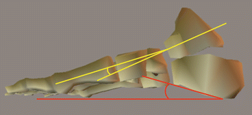
Fig. 30.2
Calcaneal pitch angle (red line) and talar-first metatarsal angle (yellow line)
Clinical evidence of a varus hindfoot was apparent in 16 ft (76.2 %), whereas radiographic measurements revealed varus alignment in 18 ft (85.7 %). The authors proposed that varus hindfoot alignment is a risk factor for the development of Jones fracture.
Lee et al. (2011) reported radiographic data from 50 consecutive athletes with a diagnosis of fifth metatarsal stress fracture (case group). Relative to the control group, the case group exhibited a significantly larger talocalcaneal angle and calcaneal pitch on lateral radiographs (Fig. 30.3).
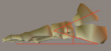
Fig. 30.3
Calcaneal pitch angle and talocalcaneal angle
On the anteroposterior radiographic view, the case group exhibited significantly greater lateral deviation of the fifth metatarsal (Fig. 30.4a) and a significantly smaller fourth-fifth intermetatarsal angle (Fig. 30.4b). The authors concluded that a large talocalcaneal angle and calcaneal pitch angle, representing a cavus foot, and increased curvature of the fifth metatarsal were associated with fifth metatarsal stress fracture.
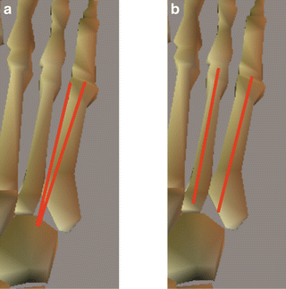
Fig. 30.4
(a) Fifth metatarsal lateral deviation angle (left) and (b) fourth-fifth intermetatarsal angle (right)
Yoho et al. (2012) reported a relationship between a transverse plane forefoot and Jones fracture. The forefoot adduction angle in the transverse plane was assessed from anteroposterior radiographs. The Jones fracture group exhibited a significantly larger forefoot adductus angle than the control group. Another study also measured the radiographic forefoot adductus angle in patients with stress fractures of the fifth and fourth metatarsal bones (Theodorou et al. 1999). The author noted that patients with metatarsus adductus are overrepresented among those with stress fractures of the lateral metatarsal bone. Therefore, forefoot adductus may contribute to the risk for Jones fracture and stress fracture of the fifth metatarsal.
Related posts:
 Injury and Illness Surveillance Among Olympic Athletes: Summary of the 2010 Winter, and the 2008 and 2012 Summer Olympic Games
 Anterior Cruciate Ligament Injury Prevention in Female Adolescents
 Anterior Cruciate Ligament Injury Prevention in Female Lacrosse Players Based on an Examination of Knee and Hip Joint Mechanics During Drop Vertical Jumps Performed While Holding a Lacrosse Stick
 Kinematics of the Foot and Ankle
 Prevention of Head and Neck Trauma in Rugby
 Electromyographic Analysis of Deep Trunk Muscles During Sports Activities
Stay updated, free articles. Join our Telegram channel

Full access? Get Clinical Tree


