Fig. 23.1
PTA after previous both column plating of an acetabular fracture
Bone Stock
Patients with PTA from acetabular fractures treated nonoperatively may have altered bony anatomy secondary to remodeling and may have deficient bone stock that requires bone graft [3]. For preoperative planning, a computed tomography (CT) of the hip can be performed to define bony morphology, and acetabular bony deficiencies can be assessed using the Paprosky classification [4]. This classification system is diagnostic and prognostic based on the location and size of the bony defects. Type I defects have a supportive bony rim with no bone lysis or migration and can often be treated by primary THA. Type II defects have intact bony columns but have distorted acetabular sockets with <2 cm of migration. These defects may be treated with autograft, allograft, or metal augments. Type III defects have severe ischial and medial osteolysis with more than 2 cm of superior migration, and these defects may require more extensive acetabular reconstruction using structural grafts, mesh, jumbo cups, or cages.
To reconstruct these defects, the goal is to create a concentric bone bed for acetabular cup placement, and it is best to place the cup in contact with native healthy bone to provide bony ingrowth. It is important to ensure that the operating room has enough bone allograft available, including demineralized bone matrix, cancellous chips, and larger structural allograft. For a primary THA, the femoral head can be used as autograft. If the defect is contained, bone graft can be placed in the defect and reversed reamed or impaction grafting can be used [5]. If there is evidence of protrusion, wire mesh or structural grafting using allograft or autograft can be fixed to the pelvis and used to add support to the acetabular cup [6]. Finally, if bony support is inadequate, trabecular metal augments of various shapes and sizes can be used to fill structural defects [7]. These augments are fixed to the pelvis with screws and cemented to the acetabular cup, as demonstrated in Fig. 23.2.
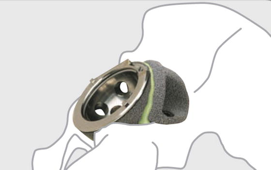
Fig. 23.2
Trabecular metal augments are used to fill bony defects and provide structural support to the acetabular cup (Printed with permission from Zimmer, Warsaw, IN)
Acetabular Fixation
In the past, acetabular cups were commonly cemented in if performed after acetabular fractures. Cups that were cemented in the acetabulum after impaction grafting when performing THA after acetabular fractures demonstrated 100 % survival at 10 years and 80 % survival at 15 years [8]. Currently, uncemented acetabular cups have become more popular as the metal surfaces of implants have improved. Titanium porous-coated sockets have shown good 10–16-year survival when performed in patients who developed PTA from previous acetabular fractures [9], and patients with trabecular metal acetabular components were found to have adequate fixation in native bone with less than 50 % contact at a minimum of 2-year follow-up [10].
When using uncemented components, there are multiple options for fixation. Multihole cups provide more screw options in the dome for acetabular cup fixation, which can provide greater support if press fit fixation cannot be achieved in the acetabulum. Screw fixation in the acetabular rim of components may provide additional fixation, but these thicker shells may reduce head size and liner thickness by 6 mm for each corresponding cup size.
If it is difficult to achieve adequate acetabular bone fixation, a gap cup can be used if there is pelvic discontinuity. Bone graft can be placed into the acetabulum and a gap cup is temporarily applied to allow for bone graft healing with a cemented liner until definitive THA is performed with an improved bone bed. Gap cups have plate extensions that allow for fixation into the ischium, teardrop, or obturator foramen with screws, hooks, or a blade plate. However, studies have demonstrated high rates of fatigue and catastrophic failure ranging from 37 to 42 % [11, 12].
Another option is to use a cup-cage construct, where a second-generation porous titanium cup is impacted into bone (Fig. 23.3). If there is inadequate bony fixation, bone graft can be applied and an acetabular cage can be placed on top of the cup and fixed to the ischium and ilium with screws [13]. A polyethylene liner is then cemented into the cup-cage construct, and the acetabular cup has been shown to achieve osseointegration in 88.5 % of patients with no signs of loosening at 44.6 months [14].
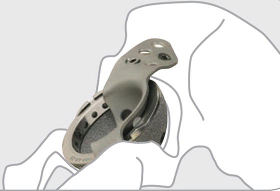
Fig. 23.3
A cup-cage construct may be used to provide fixation in inadequate bone stock by allowing the porous cup to ingrow into bone graft and native bone (Printed with permission from Zimmer, Warsaw, IN)
Intraoperative Complications
It is important to obtain adequate exposure intraoperatively to remove existing implants, place new implants, and remove HO. Despite careful surgical technique, delayed THA for PTA may have greater intraoperative complications compared to THA for OA. Heterotopic ossification (HO) is more likely to develop in patients who undergo ORIF for previous acetabular fractures, and extensive Brooker grade 3 ossification may make it more difficult to mobilize the femur. Brooker grade 4 HO, or autofusion of the hip, may require aggressive removal of HO prior to performing THA. Additionally, cutting the femoral neck in situ and performing extensive capsular release may need to be done to facilitate dislocation.
In addition to HO, patients with PTA may have significant limb length discrepancies. Scar tissue may develop after previous ORIF and may result in increased traction on the sciatic nerve. Thus, careful dissection of scar tissue is important, and using somatosensory evoked potentials (SSEPs) may be helpful for monitoring potential nerve damage, especially with leg length discrepancies greater than 2 cm.
Postoperative Management
PTA patients who undergo THA may require different postoperative management, although most patients can be managed similarly to patients with OA. If extensive bone grafting is used, then weight bearing status might need to be altered to be partial or touchdown weight bearing to reduce the forces across the hip. Weight bearing restrictions may need to be implemented for 6–8 weeks to allow for adequate healing of the acetabular implants to native and graft bone.
Additionally, patients who receive THA with PTA may have increased dislocation risk and hip precautions may need to be instituted. Avoidance of excessive flexion, adduction, and internal rotation in patients with a posterior approach to the hip may reduce dislocations. Finally, patients with previous excessive HO may require irradiation or treatment with indomethacin to prevent recurrence of HO, which can limit motion and function after THA.
Outcomes
The average time that PTA patients undergo THA after sustaining acetabular fractures ranges from 36 months to 15 years [15–19]. THA performed for PTA have generally fair outcomes, which depend on minimizing complications, such as infection and nonanatomic restoration of the hip center, as well as achieving adequate bony fixation of THA components [18, 20, 21]. A study conducted in 1978 by Boardman and Charnley found that patients that received a cemented Charnley THA after injury to the hip demonstrated good to excellent results after 15-year follow-up [16], but did not compare it to a cohort of patients without hip injury. Uncemented acetabular components have also demonstrated good outcomes, as patients had improved Harris Hip Scores (HHS) with a 5-year survival of 79 %, which increased to 97 % if survival for aseptic acetabular loosening was only evaluated [18]. While other studies have also documented improvement in HHS, these same studies have also demonstrated that performing THA after ORIF of acetabular fractures, especially those with complex fracture patterns, has increased surgical duration, blood loss, transfusion rates, sciatic nerve injuries, heterotopic ossification, and greater instability that may require treatment with an elevated acetabular liner [17, 22]. Performing a THA conversion from previous cephalomedullary nail or sliding hip screw fixation has higher complication rates, greater blood loss, and longer operative time than primary THA [23, 24]. When comparing the outcomes of THA for patients with PTA to those with OA, one study found that the patient populations had similar rates of femoral component loosing and revision, but there was four to five times greater acetabular component revision and loosening in patients undergoing THA for PTA [19].
Besides THA, outcomes of arthroplasty performed for PTA are generally poorer than arthroplasty performed for other forms of arthritis. For total knee arthroplasty (TKA) performed for PTA, there are higher complications such as extensor mechanism avulsions, increased infections, and wound breakdowns as well as poorer outcomes such as stiffness and greater instability when compared to TKA performed for OA [25–29]. Patients with total ankle arthroplasties (TAA) performed for PTA similarly had higher complication rates compared to TAA patients with OA, and PTA ankle patients also had more operative procedures [30]. For patients undergoing total shoulder arthroplasties (TSAs), patients who had the diagnosis of primary OA had longer 10-year survival compared to those diagnosed with previous fracture (94.2 % vs. 76.8 %), lower complications (primary OA 8.9 % vs. PTA 24.7 %), and higher outcome scores as determined by the Constant-Murley score (primary OA 93.7 % vs. PTA 62.7 %) [31, 32]. Patients with rheumatoid arthritis (RA) undergoing total elbow arthroplasties (TEAs) had lower outcomes scores, as measured by the Mayo Elbow Performance Score (MEPS), than for TEAs performed in patients with PTA [33, 34]. Thus, patients with the diagnosis of PTA who are undergoing arthroplasty for hips, knees, ankles, shoulders, and elbows may have poorer outcomes than patients who have primary OA or RA.
Related posts:
 Arthritis That Develops After Joint Injury: Is It Post-Traumatic Arthritis or Post-Traumatic Osteoarthritis?
Post-Traumatic Arthritis: Definitions and Burden of Disease
Potential Targets for Pharmacologic Therapies for Prevention of PTA
Arthritis That Develops After Joint Injury: Is It Post-Traumatic Arthritis or Post-Traumatic Osteoarthritis?
Post-Traumatic Arthritis: Definitions and Burden of Disease
Potential Targets for Pharmacologic Therapies for Prevention of PTA
 Outcomes of ACL Injury: The MOON Consortium
 Biomarkers of PTA
 Instability: Dynamic Loading Models
Stay updated, free articles. Join our Telegram channel

Full access? Get Clinical Tree


