Repair of Acute and Chronic Quadriceps Tendon Ruptures
Krishna Mallik
DEFINITION
Quadriceps tendon ruptures result in disruption of the fibers of this tendon, thereby disrupting the extensor mechanism of the knee.
Injury is prevalent in patients older than 40 years old and is more common in men.
Ruptures usually occur transversely through the tendon at a pathologic area approximately 2 cm proximal to the superior pole of the patella, and then progress obliquely into the medial and lateral retinacula based on the amount and duration of force.
Ruptures can occur at the bone-tendon interface (older patients) or at the midtendinous or musculotendinous area (younger patients).10
Unilateral ruptures are more common; bilateral ruptures may occur due to a predisposition from an underlying systemic condition.
Acute repair of the tendon provides a higher rate of return of function.
ANATOMY
The quadriceps tendon consists of the coalescence of the rectus femoris, vastus medialis, vastus lateralis, and vastus intermedius, 3 to 5 cm proximal to the patella, and inserts into the superior pole of the patella.
The quadriceps tendon averages 8 mm in thickness and 35 mm in width.13
Normal quadriceps tendon layers include three layers:
Superficial layer, which originates from the posterior fascia of the rectus femoris.
Deep layer, which originates from the anterior fascia of the vastus intermedius.
Middle layer, which originates from the deep fascia separating the vastus medialis and lateralis from the vastus intermedius.13
The tendon receives its blood supply from multiple contributions: branches of the lateral circumflex femoral artery, the descending geniculate artery, and the medial and lateral superior geniculate arteries.9
The distribution of the blood supply in the tendon is asymmetric.9
The superficial tendon is well vascularized from the musculotendinous junction to the patella.
The deep portion of the tendon has an oval avascular area.
PATHOGENESIS
Quadriceps tendon rupture typically occurs through a site of pathologic degeneration in the tendon caused by repetitive microtrauma.3,4
Rupture is the result of eccentric contraction of the extensor mechanism against a sudden load of body weight with the foot planted and the knee flexed.8
Rupture can be due to trauma, use of corticosteroids, and systemic diseases (gout, pseudogout, systemic lupus erythematosus, rheumatoid arthritis, renal failure, hyperparathyroidism, diabetes mellitus).5
Fluoroquinolone antibiotics (ciprofloxacin) have also contributed to tendon weakness.
Prolonged immobilization weakens the tendon, thereby increasing risk of rupture.
Although rare, ruptures can occur following total knee arthroplasty or aggressive release of lateral retinaculum.
Bilateral ruptures typically are the result of systemic medical conditions.
NATURAL HISTORY
Unrepaired quadriceps tendon rupture can lead to chronic extensor lag and weakness.
Long-term rupture may lead to quadriceps fibrosis as well as patella baja.
Partial tears can be treated nonoperatively based on the integrity of the extensor mechanism.
HISTORY AND PHYSICAL FINDINGS
Immediate pain, occasional swelling, subcutaneous hematoma
Occasionally hears or feels a “pop”
Inability to bear weight
Sensation of knee “giving away” or buckling
Preexisting pain and symptoms related to quadriceps tendon (tendinosis) prior to injury
Effusion can be indicative of hemarthrosis.
Loss of extension (straight-leg raise) indicates lack of continuity of the extensor mechanism (Note: ability to extend knee with a tendon rupture may be due to intact retinacula).
Suprapatellar gap (a soft tissue defect proximal to the superior pole of the patella) is indicated by loss of continuity of the extensor mechanism at the quadriceps tendon attachment.
Patella baja (patella of the injured knee more inferior than the contralateral knee) is indicated by loss of proximal extensor mechanism.2
Incomplete rupture—knee may extend when fully supine, however unable to extend from a flexed position
Chronic rupture—easily missed
Difficulty ambulating
Pain—may be nonspecific, typically anterior knee
IMAGING AND OTHER DIAGNOSTIC STUDIES
Plain radiographs (especially lateral view) may demonstrate bony avulsion fractures at the superior patella or soft tissue calcific depositions in chronic tendinosis.
Tooth sign6: on Merchant view, vertical ridging of osteophytes at the quadriceps tendon attachment site
Ultrasound, although operator dependent and not as specific, may demonstrate a discrete break in the tendon with abnormal overlying soft tissue.
Arthrography is invasive; however, it is positive with extravasation of contrast dye from the suprapatellar pouch and along the sheath of the tendon.1
Magnetic resonance imaging (MRI) remains the gold standard in diagnosing partial and complete quadriceps tendon ruptures in addition to associated soft tissue injuries.
Notable findings include focal tendon discontinuity, increased signal in the tendon, wavy patella tendon as well as possible preexisting pathology.
DIFFERENTIAL DIAGNOSIS
Patella tendon rupture
Quadriceps tendon rupture
Patella femoral contusion
Cartilage contusion
Neural injury
Patellar fracture
NONOPERATIVE MANAGEMENT
Patients with partial quadriceps tear, but functionally intact extensor mechanism, may be treated nonoperatively.
For the first 6 weeks, immobilize knee in extension to assist with tendon healing and maintenance of tendon length.
This can be done with a long-leg brace locked in extension or with a long-leg cylinder cast.
Patients should initially be non-weight bearing with crutches.
Patient may begin isometric straight-leg raises.
In the next phase, regaining flexion is emphasized, and the brace is unlocked to allow restoration of normal gait.
The patient is advanced to full weight bearing once stable range of motion is demonstrated.
In the last phase, strengthening is emphasized.
Patients can return to activity once full range of motion and strength are restored, typically in 4 months.
SURGICAL MANAGEMENT
All complete tendon ruptures should be repaired acutely to restore extensor function.
Any partial rupture that has progressed to a complete rupture should also be repaired as soon as diagnosed.
Preoperative Planning
Review all imaging studies.
Confirm any associated injuries that will require surgical attention.
Early treatment decreases risk of tendon scarring and loss of tissue excursion.
Chronic injury may require additional allograft tissue for reconstruction.
Positioning
The patient should be placed supine on the operating table with all bony prominences padded.
A bump under the ipsilateral hip can prevent external rotation of the operative leg.
If an examination under anesthesia is necessary, care must be taken not to convert a partial tear to a complete rupture.
Avoid use of tourniquet as this may inhibit excursion of tendon tissue.
Approach
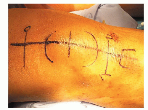
A midline patella incision, centering over the bone-tendon interface, provides access to the tendon repair in addition to evaluation and repair of the medial and lateral retinacula (FIG 1).
TECHNIQUES
▪ Acute Tendon Repair at the Tendon-Bone Interface
Tendon Preparation
A straight, full-thickness, 10-cm midline incision is made, centered over the bone-tendon interface.
Retract superficial layers to examine the deep tissue layers (TECH FIG 1A).
Irrigate hematoma.
Evaluate medial and lateral retinacula (TECH FIG 1B).
Stay updated, free articles. Join our Telegram channel

Full access? Get Clinical Tree


