Modalities and exercise
Time commenced
Early rehabilitation
Immobilization in ER
Immediate
NSAID
Scapular isometric setting exercises, addressing excessive rotation in any of the three cardinal planes
Wrist, hand, and elbow supported active range of motion to maintain function
Remote trunk and lower limb kinetic chain exercise to address individual functional deficits (e.g., leg extension)
Cardiovascular maintenance exercise (e.g., stationary cycling) – after third day to ensure no further bleeding is caused
Optimizing scapulohumeral and kinetic chain integrated function
(A) Gradually restore full range of pain-free movement
3 weeks
Active and active assisted exercises, manual therapy
(B) Scapulohumeral function
Rotary glenohumeral joint control in increasing elevation and under load (e.g., isometric progressing to isotonic ER and IR) ensuring minimal displacement of axis of GH rotation
Weight-bearing exercises (e.g., four point kneeling, push-up plus)
Glenohumeral joint elevation under load
Global shoulder function – pushing and pulling (e.g., chest press, rowing)
(C) Kinetic chain function
Strengthening of the elbow, wrist, and hand
Integrated kinetic chain exercises (e.g., PNF D2 pattern, plank exercises)
Sports specific kinetic chain
Surfing examples
Trunk extensor strength (paddling position)
Prone trunk extension and glenohumeral ER on fitball (Fig. 17.1a)
Kayaking examples
Unstable sitting with glenohumeral rotation (Fig. 17.1c)
Power and sport-specific function
Surfing examples
12+ weeks
Paddling with resistance and increasing speed (Fig. 17.1b)
Pop-ups
Kayaking examples
Swiss ball cable chop (Fig. 17.1d, e)
Isometric holds in abduction, in rotation with repeated body drop to mimic injury mechanism
From approximately 3 weeks, the goal of rehabilitation is to restore full range of motion and coordinated and sport-specific function of scapular and glenohumeral muscles with the entire kinetic chain (Table 17.1). The shoulder is inherently unstable, so aside from the passive stabilizing structures (e.g., glenohumeral capsule, ligaments, labrum), active stabilizers, primarily the rotator cuff, are essential for joint stability during function [20, 21]. Rehabilitation is tailored to the individual athlete, for example, addressing underlying glenohumeral internal rotation deficit (Table 17.1, A). It is important to reestablish rotator and elevation function under load while maintaining a stable glenohumeral joint and with minimal or no symptoms (Table 17.1, B). The scapula provides a stable base for shoulder function and is a key link in transfer of power from the lower limbs and trunk to the arm during sporting function. Weight-bearing exercises stimulate proprioception and encourage co-contraction of scapular and rotator cuff muscles [22, 23] (Table 17.1, B). Activation of subscapularis is important, but overactivity in pectoralis major should be avoided [17]. Muscular balance in pushing and pulling is important in kayaking [24], and global muscle exercises including rowing and chest and shoulder press should be incorporated, although machine pectoralis major exercises in shoulder abduction and external rotation (e.g., “pec deck”) should be avoided.
Sport-specific kinetic chain function exercises are designed to replicate the strength and endurance demands of the trunk, shoulder girdle, and glenohumeral joint during sport (Table 17.1, C and Fig. 17.1). The final rehabilitation phase involves introduction of power (force times velocity) and replicating sport-specific challenges to the glenohumeral joint (Table 17.1 and Fig. 17.1). Speed should not be commenced until single-arm loaded shoulder rotation and elevation strength is a minimum of 80 % of the unaffected side and can be performed with minimal or no pain.
Fig. 17.1
Sport-specific surfing exercises: (a) glenohumeral external rotation in trunk extension on fitball, (b) simulated paddling with glenohumeral control with increasing speed. Sport-specific kayaking exercises: (c) glenohumeral external rotation during unstable sitting on fitball, (d, e) cable chop on fitball – right glenohumeral relative abduction and external rotation in initial position
Return to Sport
The most important principle of safe return to extreme sport following shoulder dislocation is reducing the risk of recurrence by modifying sporting activity in the short term. This includes avoiding kayaking and surfing in rough water and avoiding challenging and wet snow slopes that increase the risk of falling. Returning to kayaking is particularly challenging as the lower limbs and trunk are fixed and contribute less to absorption of rotator forces, and abduction and external rotation of the shoulder under load are characteristic of common kayaking maneuvers, including the brace and roll. This highlights the necessity for tailored sport-specific rehabilitation replicating the demands placed upon the shoulder in kayaking prior to full return to sport. Athletes may return to sport with accelerated rehabilitation program by 4 months [25], although this may be longer depending on the intensity of sports (e.g., white water kayaking). Prior to return to sport, any technique faults need to be identified and addressed by developing strength and flexibility or modifying high-risk positions.
A2 Pulley Rupture
Finger injuries are the most common upper limb region injured in climbers, and A2 and A4 finger pulley injuries (climber’s finger) the most common climbing injury [26, 27], particularly in sports compared with traditional climbers [27]. Briefly, several retinacular thickenings function as pulleys for the flexor digitorum superficialis and profundus tendons. Please see Chap. 2 for further description of A2 pulley injury (Figs. 2.7, 2.8 and 2.10, pages 22–23).
The most common mechanism involves moving quickly from a crimp to a more open grip, often due the foot slipping from a foot hold [28, 29]. During this movement, the flexor tendons undergo eccentric breaking at speed. The force in the tendon may be lower in this eccentric movement compared to a concentric movement, probably due to friction between the pulley and tendon that increases risk of pulley injury [30–32].
Partial ruptures of the A2 or A4 pulley may not present with clinically obvious bowstringing, whereas this is a feature of complete A2 and A4 pulley rupture [33]. Pain and swelling is usually localized to the base of the proximal phalanx and is aggravated by flexion, especially when resisted. There is often a loss of power in gripping functions, most likely related to reduced moment arm of the flexor tendons, but may also be related to pain inhibition in the acute phase.
Treatment Modalities and Rehabilitation
The treatment of pulley injuries depends on the extent of pathology (assessed accurately with ultrasound or MRI), and this has been fully described by Schoffl et al. [28]. A strain or partial rupture may not require any immobilization, although the fingers are taped for pulley protection. Complete pulley ruptures are immobilized for up to 2 weeks with a hand-based resting splint in a functional position (Fig. 17.2a) that minimizes forces at the A2 pulley by preventing metacarpophalangeal (MCP) flexion. Activities involving MCP joint flexion increase A2 pulley load and should be avoided. In the acute phase, nonsteroidal anti-inflammatory drugs (NSAID) are recommended, although this depends on the extent of swelling and pain. Soft tissue therapy and ultrasound to reduce swelling and encourage healing are indicated.
Fig. 17.2
Rehabilitation of A2 pulley injuries: (a) immobilization in resting splint, (b) MCP joint block ring splint, (c) resisted isolated fourth MCP flexion, (d) simulated crimp grip with putty resistance, (e) resisted MCP and PIP flexion with DIP extension, (f) grip strength measurement with handheld dynamometer
At between 2 (partial rupture) and 2–4 weeks (complete rupture), functional therapy including range of motion exercise is commenced, followed by progressive loading of the flexor tendons. An MCP joint block ring splint (Fig. 17.2b) can be used for ongoing protection of the A2 pulley. Active (unloaded) exercise is progressively replaced with resistance (loaded) exercise (Fig. 17.2c). Gradual increase of forces acting at the A2 pulley during the rehabilitation phase is paramount to facilitate pseudo-pulley formation. Consequently, loaded MCP joint flexed positions and “crimp” positions are introduced in the later stages of recovery (Fig. 17.2d, e). As exercises are progressed, finger pulleys can be protected during rehabilitation with circular tape or a thermoplastic ring. Schweizer [34] demonstrated a biomechanical advantage in reducing the extent of flexor tendon bowstringing by taping the distal aspect of the proximal phalanx instead of over the A2 pulley.
Finger pulley injuries often occur at the end of a climb [35] and are more common in sports climbers who perform more challenging ballistic movements; this highlights the importance of adequate muscular strength rehabilitation in both the upper and lower limb kinetic chains. General grip strength can be measured with dynamometer (Fig. 17.2f). Using the first rung position of the Jamar dynamometer will enable measurement of grip in an intrinsic (lumbricals and interossei) dominant position of MCP flexion – the adopted posture for the “crimp” climbing position. In this way, response to treatment can be objectively monitored as can readiness for a return to climbing activities.
Return to Sport
Once grip strength is a minimum of 80 % of the unaffected side, climbing activity can commence. This is usually a minimum of 4 weeks for a partial rupture, 6 weeks for complete rupture, and 4 months postsurgical repair [28]. Crimp grip should be avoided for a further 6–8 weeks following complete A2 pulley ruptures and surgical repair [36], given the much larger A2 pulley forces with this grip [37]. Strength criteria and careful symptom monitoring as progressing too quickly are common and are often associated with overconfidence of the climber in attempting difficult climbs too early. Circular tape as described above is commonly used by climbers, but cadaver evidence suggests it may not increase the maximum load at failure that the pulley can sustain [38], whereas other studies have found that taping is minimally effective in absorbing flexor tendon bowstringing forces [34].
Anterior Cruciate Ligament Rupture
Anterior cruciate ligament rupture is common in skiing and less so in snowboarding [5]. The mechanism of injury often involves external knee rotation and valgus (lateral to medial) loading while turning and catching the inside edge of the downhill ski or an unbalanced landing from a jump in higher level skiers [39]. Regardless of the level of the athlete, they can expect to return to sport after management, usually involving surgical repair (ACLr) followed by prolonged rehabilitation for higher level skiers. Nonsurgical management may be suitable for skiers at all levels [40, 41], and conservative management success is predicted by a lack of concomitant ligament or meniscal injury, reasonable function (e.g., performance on hopping tests), and minimal or no giving way [42]. There is an emphasis on criteria-based progression through a structured rehabilitation program with both nonsurgical and surgical management, and this facilitates a timely and safe return to sport.
Treatment Modalities and Rehabilitation
Generic post-ACLr rehabilitation has been described in detail elsewhere [43, 44]. In this section, a suitable rehabilitation program for skiers and snowboarders will be outlined with reference to sport-specific exercises and progression criteria. The early rehabilitation phase (0–4 weeks) focuses on managing surgical pain and swelling with ice, compression, elevation, relative immobilization, soft tissue therapy and muscle recruitment strategies, and critically, reestablishing pre-injury level of knee extension and flexion. Core and trunk strength exercise and rehabilitation of specific kinetic chain deficits can commence. Bracing post ACLr is a common practice; however, a recent review concluded that it may not influence re-injury rate and function at long-term follow-up [45]. Decisions regarding duration of brace use are based on athlete confidence, adequacy of muscle control during activities of daily living, and the stability of secondary knee restraints.
In the second phase (5–16 weeks), the focus is on muscle strength, hypertrophy, and power, as well as neuromuscular control flexibility. The emphasis is on single-leg exercise to address lower limb kinetic chain asymmetry and should focus on quadriceps and hamstring strength and hypertrophy as well as strength in the distal (calf) and proximal (gluteal, hip flexors) kinetic chain. There is an ongoing debate about the relative efficacy of closed- versus open-chain exercises. Tagesson et al. [46] built on the work of Morrissey et al. [47] showing no difference in laxity, pain, hamstring strength, jump performance, and functional outcome (Knee Injury and Osteoarthritis Outcome Score) between open- and closed-chain rehabilitation. The only difference was greater quadriceps strength in the open-chain group, suggesting that open-chain exercise should be considered in ACLr rehabilitation programs. It is therefore an athlete-specific decision regarding how to balance open- and closed-chain exercise depending on sport-specific requirements. Depending on the graft, there needs to be a focus on restoring quadriceps strength (patellar tendon graft) or hamstring strength (hamstring graft) [48].
Afferent proprioceptive input moderates neuromuscular control in both feedforward and feedback mechanisms to allow efficient and coordinated athletic function. In the second rehabilitation phase, this involves balance, nonimpact ski-specific exercises (e.g., Fig. 17.3), and unexpected support surface perturbation exercises. In the third phase of rehabilitation (17–22 weeks), the emphasis is on advanced neuromuscular exercise involving impact. This includes agility, plyometric, running, and sport-specific training. The addition of neuromuscular training to conventional resistance training may improve pain and functional scores [49, 50]. There is an emphasis on reproducing sport-specific lateral knee movements under increasingly challenging conditions.
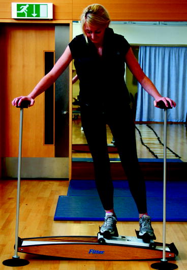
Fig. 17.3
Ski-specific neuromuscular rehabilitation on a “fitter”
Return to Sport
The fourth and final phase of ACLr rehabilitation involves return to play. Important return to sport criteria include a lack of functional instability or giving way, strength, power, functional outcome score within 10 % of opposite side, and successful completion of agility, plyometric (e.g., crossover triple hop), and sport-specific drills and training [44, 51]. Skiers who wore a functional knee brace when returning to sport were found to have a 2.74 times lower risk of knee injury [52]. Adequate warm-up is the keystone of ACL prevention and should involve a neuromuscular warm-up addressing trunk and lower limb control and coordination in functional tasks [53]. Several warm-up programs (e.g., “the PEP,” “the 11+”) including balance, strength, plyometric, and agility exercises have been shown to reduce the risk of first-time ACL injury [54–56]. Factors that increase the propensity for the knee to move into a valgus pattern in single-leg squatting and landing should be addressed, particularly in females among whom this pattern is recognized [57].
Overuse Injuries
Wrist Extensor Tenosynovitis and Intersection Syndrome
Tenosynovitis of the distal extensor tendons of the wrist is common in kayakers/canoeists as well as other sports (rowing, racquet sports). In long distance kayaking, the incidence of overuse injuries to the wrist is between 21 and 50 % [58, 59]. Intersection syndrome is commonly reported in kayakers and alpine skiers and occurs where the tendons of the first extensor compartment cross over the tendons of the second extensor compartment 4–5 cm proximal to Lister’s tubercle (Fig. 17.4). Palmer et al. [60] reported a 12 % prevalence of intersection syndrome among alpine skiers. The etiology of intersection syndrome may involve friction between the first and second compartment extensors [61] and hypertrophy of the APL and EPB tendons [62].
Fig. 17.4
Intersection syndrome symptoms are localized to the crossover between the first and second extensor compartment tendons, 4–6 cm proximal to Lister’s tubercle
The mechanism of both injuries is repetitive excessive wrist extension, for example, during the pull phase in kayaking. Among kayakers, injury is more common in longer stages (>38 km), rougher water, or kayaking on multiple consecutive days, and greater training prior to events is preventative [59]. Weakness and inefficient use of the proximal upper limb kinetic chain and trunk may also increase wrist overuse and lead to injury. The underlying pathology is inflammatory, with swelling and crepitus often evident in the distal and radial aspect of the dorsal forearm.
Treatment Modalities and Rehabilitation
The key element of management is reducing repetitive loads and correcting technique faults predisposing to injury. This includes addressing functional deficits in the entire upper limb kinetic chain and trunk, for example, reduced trunk strength that may increase wrist extension when the kayaker is pulling. Complete rest from kayaking and skiing is often required initially, although partial rest may be sufficient in less severe cases. If symptoms do not settle, immobilization in a static wrist splint (prefabricated thermoplastic wrist splint in 15° extension) for 2 weeks is indicated [63]. Conservative treatment includes ice, distal to proximal massage (fingers to elbow), stretching the wrist extensors, crepe bandage to encourage reduction in swelling worn at night or times of activity [64]. Wrist extensor stretching should not be performed if painful as this may indicate further compression and injury to the tendon sheath [3]. Ultrasound-guided cortisone injection into the extensor tendon common sheath is indicated if symptoms do not settle within 1–2 weeks following immobilization/load modification [65]. There are currently no outcome studies comparing the influence of platelet-rich plasma in tenosynovitis; however, this is an area which may benefit from this new modality.
Return to Sport
The risk of tenosynovitis may increase during multiple day events [59]; therefore, it is important when returning to kayaking to allow sufficient rest time between endurance training sessions (48–72 h initially). Modification of equipment may help to relieve symptoms, such as changing the diameter of ski poles among skiers [66] that may improve the length-tension relationship of the wrist extensors.
Ulnar Nerve Neuropathy
Compression of the ulnar nerve in Guyon’s canal (cyclists palsy) is common in long distance cycling, including mountain biking [67]. Guyon’s canal is formed by the pisiform medially and hook of hamate laterally. The motor branch of the ulnar nerve to the hypothenar muscles is given off before Guyon’s canal, and compression at this site leads to weakness of all ulnar-innervated hand muscles (hypothenar, interossei, third and fourth lumbricals) and skin sensation. Compression at Guyon’s canal or distally is associated with only sensory loss and/or intrinsic muscle weakness (interossei, third and fourth lumbricals and adductor pollicis), depending on whether the superficial sensory or deep motor branches are compressed [68]. Differential diagnosis includes stress fracture of the hook of hamate.
Patterson et al. [69] assessed ulnar nerve sensory function among 18 mountain bike and 32 road bike riders before and after a 600-km ride. Thirty-nine percent had signs of ulnar nerve motor deficit and 33 % and had sensory loss; mountain bikers were more likely to report sensory loss than road bikers. Risk factors include long distance cycling, asymmetrical weight bearing on the handlebars, poor core stability, poor seat or handle bar position encouraging wrist hyperextension, and inadequate handlebar and glove padding [69–71].
Management
The most important aspect of management is to minimize compression of the ulnar nerve during cycling. This can often be achieved by adjusting the mountain bike riders grip and hand position on the handlebars, padded handlebars, and gloves and frequently changing hand position while mountain biking [72]. Padded gloves have been shown to reduce hypothenar pressure in cycling by between 10 and 28 % [73]. If symptoms persist, it is important to progressively reduce the volume of mountain bike riding until there is adequate resolution of symptoms. This is particularly important if the mountain bike rider is experiencing pain or weakness with daily activities such as gripping.
Management should include assessment and treatment of cervical and upper limb neurodynamic dysfunction, seeking evidence of subtle “double crush” nerve compromise. Smith et al. [74] found that cyclists with ulnar nerve compression symptoms at the wrist were more likely to have positive thoracic outlet provocative tests and concurrent neck and shoulder pain compared with matched controls. Neurodynamic dysfunction is initially addressed by working on neural interfaces such as mobilization of the shoulder and first rib, while trying to reduce nerve irritation by avoiding end of range positions, followed by more direct nerve gliding techniques (e.g., upside down goggles stretch) in progressively more provocative positions [75]. Surgical release of Guyon’s canal is uncommon but indicated in recalcitrant cases.
Extension-Related Low Back Pain
Low back pain (LBP) is common among sailors, surfers, and windsurfers, often related to prolonged maintenance of end range postures or repetitive movement. This often happens in “pumping” the sails to accelerate the boat and “hiking,” which involves positioning (usually in lumbar extension) the body to prevent the boat leaning away from the wind. During surfing, hyperextension of the lumbar spine occurs during paddling out into the surf. Twelve percent of injuries in big boat sailing [76] and 22 % in windsurfing [77] may involve the lumbar spine. Extension-related LBP is also common in cross-country skiing and has been linked with increased thoracic kyphosis and lumbar lordosis [78]. The most common underlying pathology is facetal and disc spondylosis in older and pars defects in younger sailors and windsurfers, probably related to repetitive extension movement [79, 80]. Symptoms can be localized unilateral or bilateral low back pain aggravated by prolonged extension postures and standing.
Treatment Modalities and Rehabilitation
The principles of treating extension-related LBP in sailors and windsurfers are outlined in Table 17.2. Initial management involves avoidance of aggravating activities or postures, while rehabilitating motor patterns demonstrate segmental or region-specific extension /rotation overstrain and sport-specific strength. This period may be between 7 days and 3 months, depending on the severity of symptoms, underlying pathology, athlete engagement, and motor learning skill. For example, a pars defect may require 2–3 months rest from end of range extension postures (e.g., pumping, hiking) [81–83]. NSAID, ice, and kinetic chain exercises remote from the painful site are indicated in this phase. Efficient transfer of load from the lower limbs to the spine is critical in windsurfing and sailing, for example, in hiking. Quadriceps strength is an important determinant of average hiking moment produced by sailors, and this in turn predicts race scores among Laser sailors [84].
Table 17.2
Rehabilitation for extension-related LBP among sailors and windsurfers
Exercises | Time commenced | |
|---|---|---|
Early rehabilitation | Load management (e.g., sailing, excessive standing/walking) | Immediate |
Ice, NSAID | ||
Remote upper and lower limb kinetic chain exercises to address individual deficits (e.g., calf raises) | ||
Cardiovascular maintenance exercise (e.g., upper limb ergometer) | ||
Dissociation of extension from distal (hip) and proximal (shoulders) kinetic chain | ||
Optimising lumbopelvic muscle function | Lumbopelvic muscle-tendon compliance and strength | 2 weeks |
Hip flexor flexibility (tensor fascia lata, rectus femoris, sartorius) (e.g., split squats into end of range) | ||
Abdominals (e.g., crunches) | ||
Gluteals (e.g., bridging) | ||
Hip flexor strength (e.g., standing hip flexion with pulleys) | ||
Avoid back extension exercise into end of range in favour of exercises loading the back extensors isometrically (e.g., McGill birddog exercise, ref) | ||
Local and global muscle activation | ||
Kinetic chain function | ||
Integrated kinetic chain exercises (e.g., squats) | ||
Sports specific kinetic chain | ||
Front plank progressions for abdominals, hip flexors Fig. 17.5 | ||
Sport-specific exercise and graded return to sport | Simulated ‘pumping’ – pulleys | 6+ weeks |
Increasing volume and speed | ||
Graded return to windsurfing and sailing |
The second phase focuses on optimizing lumbopelvic function while maintaining movement patterns that reduce end range extension stress on previously painful structures. Improving the flexibility of pelvic muscles that maintain lumbar lordosis [85–87], including the tensor fascia lata (Table 17.2), may reduce lumbar extension moment and load on damaged spinal structures. Strong abdominal and hip flexor muscles are necessary to control lumbar spine posture in pumping [88]. They can be strengthened with progressive front plank exercises (Fig. 17.5). Compliant hip flexors allow greater combined hip and lumbar extension that may relatively off-load the spine. End range functional eccentric exercise for the hip flexors such as lunging can increase hip flexor musculotendinous compliance [89, 90]. Lumbar extensor muscle weakness is associated with low back pain in sports involving repetitive extension [91] and should be addressed with isometric loading exercises that avoid end of range. Recruitment of deep stabilizing muscles [92] has been shown to improve function and pain in spondylosis [93] but need to be progressed to include challenge to both local and global muscles systems [94]. A functional core strengthening exercises may be best suited to younger athletes with lumbar hypermobility [95, 96].