Reduction and Fixation of Lateral Condyle Fractures of the Distal Humerus
Patient Selection

Figure 1Illustrations show the Jakob classification of lateral condyle fractures. A, Type I: displaced less than 2 mm with no violation of the articular surface. B, Type II: displaced 2 mm or more with no violation of the articular surface. C, Type III: displaced 2 mm or more with disruption of the articular surface.
These fractures typically occur after falling onto outstretched hand or from a height
Jakob classification describes these fractures (Figure 1)
Treatment is based on extent of displacement, not location of fracture
Reserve nonsurgical management for nondisplaced or minimally displaced fractures (<2 mm)
Surgery is recommended for displaced (>2 mm) or rotated fractures
Closed reduction with percutaneous pinning may be indicated for minimally displaced fractures
Open reduction and internal fixation is warranted for significantly displaced (>4 mm) or unstable fragments
Preoperative Imaging
AP, lateral, and internal oblique views of elbow
Oblique view helps visualize true displacement
Often larger than visualized on plain films due to cartilaginous component
Hinging is difficult to assess on plain radiographs; MRI or magnetic resonance arthrography can help
Most treatment decisions are made from plain radiographs; advanced imaging rarely used because of cost and required patient sedation
Procedure
Special Instruments/Equipment/Implants
Fluoroscopic unit
Sterile tourniquet
Small right-angle retractors for open reduction
Kirschner wires (0.062-in); a small cannulated screw/washer can be used for older children with a bony fragment
Surgical Technique
Closed Reduction and Percutaneous Pinning
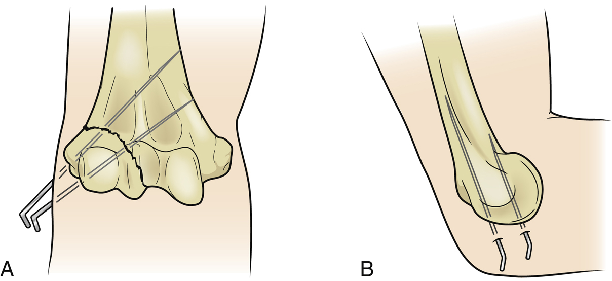
Figure 2Illustrations of the anterior (A) and lateral (B) aspects of the elbow demonstrate optimal pin placement for a lateral condyle fracture.
Stay updated, free articles. Join our Telegram channel

Full access? Get Clinical Tree


