Reconstruction of the Diabetic Charcot Foot

Surgical Reconstruction of the Diabetic Charcot Foot


Internal, External or Combined Fixation?


Keywords


• Charcot foot • Diabetes mellitus • Internal fixation • External fixation • Surgery • Reconstruction


Introduction


The causes of Charcot neuroarthropathy (CN) are numerous and multifactorial. As a result, multiple diabetic-related comorbidities are commonly encountered in patients with CN. Careful consideration and management of these comorbidities are important in determining the overall treatment strategies along with fixation methods. The presence of peripheral arterial disease is commonly seen in this diabetic population and many times can be found in the early stages of CN. However, most of the deformities associated with CN that are being considered for surgical management are chronic and long-standing. For this reason, patients with CN should be closely evaluated for peripheral vascular disease in any stage of the pathology and before any major reconstruction.


Delayed wound healing and infection are commonly the reasons for surgical failure and subsequent lower extremity amputation in diabetic patients with CN. The presence of immune dysfunction and hyperglycemia results in an impaired inflammatory process that alters the delivery of critical growth factors leading to a negative impact of wound healing. Other causes that could eventually impede wound healing are smoking, morbid obesity, malnutrition, anemia, arterial insufficiency, neuropathy, and nephropathy. A preoperative assessment to address controllable risk factors is important to improve the local environment for favorable wound healing.


Delayed bone healing related to metabolic alterations associated with diabetic fractures and/or CN is also common in this diabetic population. The exact mechanism of diabetic nonunion has yet to be determined, but the neurovascular and autonomic dysfunctions along with inflammatory mediators and metabolic derangements can lead to decreased bone callous formation in the presence of increased osteoclastic activity. The combination of poor diabetic fracture healing along with recurrent or progressive fracture patterns is a common phenomenon in diabetic patients with CN. For this reason, fixation methods must consider the delay in bone healing, decreased bone mineral density, and/or potential for the loss of fixation when reconstructing foot and ankle deformities associated with CN.


Clinical decision making for diabetic Charcot foot fixation


When evaluating a surgical patient with CN, a thorough history and physical examination are necessary to identify the duration and progression of the deformity. Clinical and radiographic findings are assessed for deformity staging, instability, or presence of reducibility. Further medical imaging such as computed tomography (CT) scan or 3-dimensional CT scan may need to be evaluated to determine the bone stock, fracture patterns, and joint malalignment. Determining the degree of bone loss or infected bone segments that may have to be surgically resected is essential in the preoperative period. Consideration of previous management is also important, especially if failed surgical attempts have been performed.


The clinical decision to recommend surgery for CN is multifactorial and patient-dependent and usually depends on the severity of the deformity with or without an ulceration or infection, presence of instability with or without preulcerative lesions that are not amenable to bracing, and presence of recurrent infections, osteomyelitis, and unsuccessful conservative treatments in the ambulatory patient.


Most common fixation methods used for the treatment of the diabetic Charcot foot include internal fixation, external fixation, and a combination of both fixations.


If the soft tissue envelope permits, internal plate or screw fixation can provide stability while correcting the associated deformities. Internal fixation can be used as compression or bridge plating to stabilize CN bone segments. The use of bone grafts with internal plate fixation can be used to address osseous defects of the midfoot, rearfoot, or ankle to optimize bone healing. External fixation is the least invasive technique and may be considered for patients who have a poor soft tissue envelope, large bone defects, severe deformities, peripheral vascular disease, and a history of previous ulcerations or infections. A multiplane circular external fixator can be used for gradual deformity correction, bone transport, in combination with internal fixation, and as a surgical off-loading device for concomitant soft tissue defects.


Intramedullary (IM) nailing has also been used to address unstable or chronic CN deformities of the rearfoot and ankle joints. In the event of infection, an IM nail should be avoided until infection or osteomyelitis is appropriately treated. Postoperative infected IM nailing in CN patients can be quite challenging and requires an extensive surgical management with local cemented antibiotics and stabilization before a clinical decision can be made to further proceed with revisional IM nailing, internal plate/screw fixation, or external fixation to avoid a potential nidus for recurrent infection.


Internal Fixation


Numerous internal fixation techniques have been described to stabilize osseous segments of CN. Bone resorption, fragmentation, and osteoporosis increase the technical demands of using internal fixation to address deformities associated with CN. Standard internal fixation techniques such as lag screws for interfragmentary compression even with standard plate fixation are often not sufficient to stabilize CN-related fractures or dislocations. Evolving techniques have focused on fixation placement along with the advent of stronger fixation plates and screws. The concept of bridge plating is to extend the fixation both proximally and distally using less affected osseous segments to achieve better screw fixation. A drawback of bridge plating is that otherwise normal joints are sacrificed to improve the stability of these internal fixation constructs. However, this concept has led to the theory of extended joint arthrodesis in CN patients with multiple fractures and dislocations. An arthrodesis is extended to include adjacent joints beyond the zone of injury to improve fixation and deformity correction while attempting to prevent further collapse. Anatomically designed plates by various industries have been developed to provide a more rigid construct to perform such procedures while considering the soft tissue envelope and plate design for deformity correction and mechanical stability.


Plantar plating for midfoot arthrodesis has been described as a technique that provides superior strength by placing the plate along the tension side of the arthrodesis site. The technique has been described to produce reliable arthrodesis of neuropathic midfoot deformities. Plantar plating may be suitable for select rocker-bottom CN midfoot deformities that have a sufficient soft tissue envelope and especially in addressing sagittal plane deformities at the navicular-cuneiform-metatarsal joints. Extending the arthrodesis to include the talo-navicular joint and the remaining tarsal joints with this technique can be quite challenging.


Medial plating techniques may be beneficial to address certain CN midfoot deformities with severe abduction or to provide stability to multiple transverse joints of the midfoot. Plate fixation in this manner is placed on the tension side of the deformity. In addition, plate fixation along the medial column allows the placement of screws to cross multiple cortices of the metatarsal and tarsal bones, improving the screw purchase and overall construct stability. Locking plates create a fixed-angle construct by attaching the screw to the plate. These devices were developed to improve fixation in osteoporotic bone, and careful consideration is needed to determine if adequate bone purchase is achieved, since the screw is rigidly attached to the plate. Other plating systems have combined locking and nonlocking abilities for certain osseous pathologies and anatomic placements.3,4


Intramedullary metatarsal screws have also been described for midfoot CN reconstruction. These large diameter screws are inserted through the metatarsal joints and extend across the midfoot into the talus or cuboid. Alternative techniques are to insert long axial screws from the talus or calcaneus into the midfoot or forefoot. The technique involves deformity correction and stabilization by placing guide wires for large cannulated screws, confirming the position of the foot and placement of the fixation under C-arm fluoroscopy. Advantages to this technique include the limited open approach for the fixation while the screws are entirely intraosseous, which limits hardware exposure. Possible complications to this technique may be due to lack of compression of the multiple arthrodesis sites, loss of purchase, hardware failure, and resulting deformity that leads to challenging revisional surgeries.


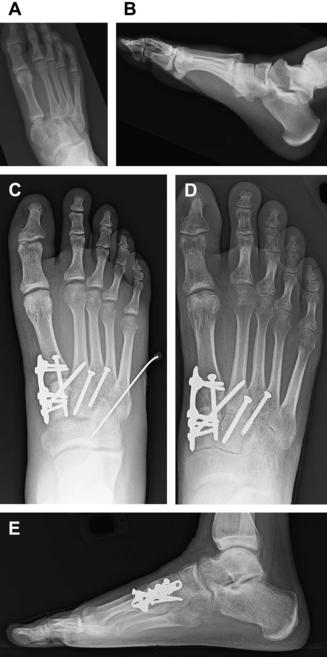
Plate fixation to stabilize the rearfoot or ankle can be used if the soft tissue envelope permits and the CN deformity if free of any ulceration or infection. Various plates and plating techniques have been described. Blade plate fixation is a fixed angle construct providing superior stability to stabilize rearfoot and ankle arthrodesis. Numerous anatomic rearfoot and ankle plating systems have been developed allowing for compression and the use of locking screws. The use of various nonanatomic plates such as the femoral and humeral plates has also been described. These plates are typically selected because of their rigidity and strength while providing multiple screw options to gain purchase into the calcaneus. Plate placement for ankle and rearfoot arthrodesis is dependent on the plane of deformity, previous surgical reconstruction, soft tissue envelope, and presence of ulceration. Lateral, anterior, and posterior approaches have all been performed. Incision placement must be carefully planned, especially if bone resections and shortening of the lower extremity are being performed, as these can result in significant tension on the surgical incisions. These clinical case scenarios need to be considered in detail before fixation constructs are placed to prevent poor soft tissue closure and postoperative complications (Fig. 1).


Stay updated, free articles. Join our Telegram channel

Mar 8, 2017 | Posted by in ORTHOPEDIC | Comments Off on Reconstruction of the Diabetic Charcot Foot

Full access? Get Clinical Tree

Get Clinical Tree app for offline access