Fig. 3.1
Action of the radioscaphocapitate ligament that can increase the displacement of the distal fragment in fractures of the scaphoid waist
This mechanism only occurs in fractures of the waist and doesn’t concern the fractures of the proximal part [8].
The same mechanism puts important constraints on the scapholunate ligament, opposed to the differential gliding between the scaphoid and lunate.
That’s why in a trauma in hyperextension, we can observe a fracture of the scaphoid, an injury of the scapholunate ligament, or a fracture of the radial epiphysis. The combination of these injuries is rare (Fig. 3.2).
Fig. 3.2
Injury mechanism that can cause a rupture of the scapholunate ligament, a fracture of the scaphoid or the radius
3.2 Clinical and Paraclinical Signs
With the patient interview only, we can determine if there has been a “risky” trauma.
The most suggestive clinical sign is pain when palpating the anatomical snuffbox, (Fig. 3.3) but if there’s an edema in this area or in the radial side of the wrist and pain when putting load on the scaphoid, we’ll have to realize X-rays [9].
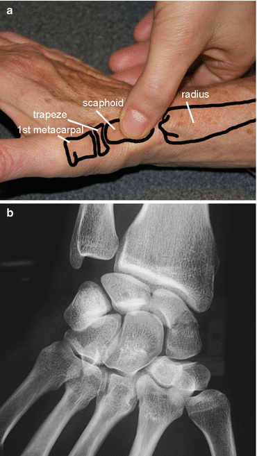
Fig. 3.3
(a) Palpation of the scaphoid in the anatomical snuffbox, between the first extensor gliding and the extensor pollicis longus. (b) Radiography showing a fracture of the scaphoid waist
Sometimes, the X-rays are clear and the diagnosis is easy, but it’s possible that the fracture isn’t visible on the X-ray on the day of the trauma.
In this case, a resting orthosis is realized and kept 15 days, and then other X-rays are realized to confirm the presence/absence of fracture.
However, we must keep in mind that the decalcification process around the fracture line that allows detecting a fracture after 15 days isn’t systematic, especially in proximal fractures. When in doubt, we use MRI and scintigraphy.
Echography also has a certain interest in the diagnosis, but its efficiency is operator dependent.
3.3 Therapeutical Process and Surgical Treatment [4–7]
The treatment depends on the location of the fracture line, whether the fracture is displaced or not, and the patient’s socioeconomical and sports context. This treatment can be orthopedic or surgical with conventional screwing, percutaneous technique, or simple pin.
3.3.1 Orthopedic Treatment [10]
The orthopedic treatment by prolonged immobilization stays the first-line indication to treat undisplaced fractures of the scaphoid, but the development of percutaneous osteosynthesis tends to progressively prevail over the cast. Several studies agree that there’s a high rate of consolidation (more than 90 %); however, opinions differ more widely as regards the type of immobilization, the wrist position, and the duration of immobilization.
3.3.1.1 Immobilize the Elbow and the Thumb?
The SOFCOT’s round table in 1988 gathers a series of 209 recent undisplaced fractures of the scaphoid treated with brachio-palmar immobilization in 41 % of the cases and antebrachio-palmar immobilization in 33 % of the cases. The 26 % left were treated with a combination of the 2 previous techniques (first brachio-palmar, then antebrachio-palmar). The conclusion of this study doesn’t bring formal statistical elements to confirm the superiority of one treatment or the other.
In 1990, a study shows on 10 subjects that pronosupination doesn’t put constraint on the scaphoid, unlike the flexion/extension and the radial and ulnar inclinations. Only two studies analyze the consolidation rate depending on whether the elbow is in the cast or not; they both conclude that the consolidation isn’t improved when the elbow is immobilized.
The fact that the elbow must be immobilized in fractures of the scaphoid is strongly anchored in people’s minds. In 1943 Watson-Jones recommends blocking the thumb’s metacarpophalangeal joint, and we have to wait until 1991 for a randomized prospective study to obtain results identical between the group of patients immobilized until the interphalangeal and those with the thumb free.
3.3.1.2 Wrist Position
The most common method is the immobilization in a neutral position in the sagittal and frontal planes, with a slight radial inclination to avoid distraction in the fracture site.
3.3.1.3 Immobilization Duration
Several studies have evaluated the consolidation duration between 12 and 15 weeks. Some authors recommend only 2 months of immobilization for distal fractures as the distal part is more vascularized, but no clinical study confirms this.
In theory, only a proof of consolidation should allow us to stop the immobilization, but in practice this consolidation is hard to confirm and immobilizing during 3 months seems to be the most scientifically established and the most prudent approach.
3.3.1.4 Immobilization Type
The cast still seems to be the most used technique. However, the resins have several advantages: They’re half as heavy as casts and they’re more solid and resist to water. But they also have disadvantages: poor plasticity making compressions more frequent and hardness of the material that can irritate the skin.
Thermoformed custom-made orthosis provides more comfort in the patients’ daily life and can be readjusted when the edema disappears.
In conclusion, 2–3 months of immobilization depending on the location of the fracture line, with the wrist in a neutral position and the thumb free in a thermoformed orthosis, is the first-line indication in undisplaced fractures of the scaphoid.
3.3.2 Surgery
Displaced fractures of the scaphoid and delayed bone healing are indications for surgery [11].
In 1968 the screwing with a spongy 4 mm screw has been described in the AO’s osteosynthesis manual, but subsequent studies have shown technique difficulties due to the particular anatomy of this bone [12] and the equipment load. Since the beginning of the 1980s and the apparition of Herbert’s headless screw, several screws have been developed to favor the compression and make the placement easier. In this chapter, we’ll develop the main osteosynthesis techniques with traditional screws, percutaneous screws, and percutaneous pins.
3.3.2.1 Traditional Screws
Screwing can be realized in a retrograde way with an anterior approach or from proximal to distal with a posterior approach in proximal fractures.
We use an AO stainless steel screw with a 2 mm diameter with a long thread length to allow a good compression (Fig. 3.4). Perforated screws avoid multiple drilling, but have a wider diameter.
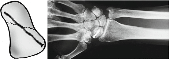
Fig. 3.4
Screw osteosynthesis
In anterograde screwing from proximal to distal, we prefer screws with a smaller diameter considering the small size of the proximal fragment (Fig. 3.5).
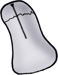
Fig. 3.5
Anterograde screwing with a small screw
The surgical follow-ups depend on the surgeon and go from no immobilization to a resin cuff during a month.
Consolidation rates vary between 90 and 100 %.
3.3.2.2 Percutaneous Screws [13]
Since a few years, little invasive surgical techniques have been developed to reduce morbidity and preserve the scaphoid’s vascularization as well as the ligaments surrounding it.
The approach can be palmar (Fig. 3.6) or dorsal, and the reduction is controlled with radioscopy or coupled with arthroscopy. This technique uses perforated screws guided by a pin. The diameter of the screw varies from 2 to 3 mm.
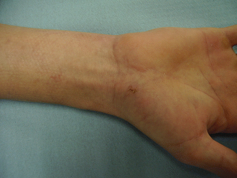
Fig. 3.6
Cutaneous aspect after percutaneous screwing
Instable and displaced fractures are formal indications for osteosynthesis [11]. The percutaneous technique is reserved to fractures that are little displaced and is more and more used in undisplaced fractures based on economic, social, professional, and sports factors, as it reduces the duration of immobilization (0–30 days).
3.3.2.3 Percutaneous Pins [14, 15]
The pins seem to be the best osteosynthesis technique for the scaphoid, putting compression on the fracture site. However, some authors are now wondering if compression is necessary for the scaphoid. The natural tendency in median fractures is towards flexion, so we’ll have to install a wiring during the synthesis. The pins allow a stable osteosynthesis without shortening of the scaphoid and avoid rotation disorders if we place at least 3 pins with a diameter between 14 and 16.
In conclusion, the surgical treatment is justified in undisplaced fractures when:
The fracture concerns the proximal part.
There’s an associated injury requiring surgery.
The fracture is open or unstable.
The patient is a sportsman or motivated worker who wants to quickly go back to his activities.
3.4 Rehabilitation and Orthotic Treatment [4–7]
Rehabilitation can follow surgery or a longer immobilization in case of orthopedic treatment.
The different protocol stages can be modified depending on the bone healing, regularly controlled with X-rays.
In any case, the priorities are regaining physiological amplitudes and strength to restore optimal function.
There are many similar points between this protocol and the one for the fractures of the inferior extremity of the radius, but the concerned patients are usually very different.
The patients with a fracture of the inferior extremity of the radius are usually woman of more than 60 years old with osteoporosis, whereas the patients with a fracture of the scaphoid are usually young men who have suffered a high kinetic energy trauma.
Therefore, the rehabilitation goals for these two pathologies are often different, as they’re related to the patient’s anterior capacities and functional imperatives. The exercises are usually more intense in fractures of the scaphoid, especially in muscular reinforcement and proprioception exercises.
3.4.1 Rehabilitation Protocol (Fig. 3.7)
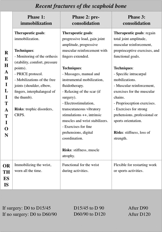
Fig. 3.7
Rehabilitation protocol
3.4.1.1 Immobilization Phase (D0 to D15/45 with Surgery or D0 to D60/90 Without Surgery)
The duration of immobilization after surgery depends on the surgical technique and varies from 15 (percutaneous screws) to 45 days (pins).
During all this phase, rehabilitation isn’t essential and self-rehabilitation advices are given to the patient.
The treatment is based on the protocol PRICE that fights against trophic disorders resulting from the trauma and surgery:
P for protection of the fracture line during the whole phase, realized immobilizing the wrist and informing the patient about the tissue fragility in this phase.
R for rest of the damaged area that mustn’t be solicited during the immobilization phase, aside from self-rehabilitation exercises explained by the therapist. These exercises are described in the Chap. 5, Fig. 5.20.
I for ice, putting cold packs several times a day on the fracture zone. If there’s a cast it’s impossible, but if there is no wound, we immobilize the patient with a thermoformed orthosis, which allows cold bath against edema.
C for compression that can be realized in the fingers if they’re swollen.
E for elevation of the wrist to avoid edema.
These exercises are combined with sensorimotor stimulations to maintain the body mapping and cutaneous receptors and with simple active mobilizations of the free joint (the metacarpophalangeals in the long fingers tend to get easily stiff). We stop these exercises if pain appears.
3.4.1.2 Pre-consolidation Phase (D15/45 to D90 with Surgery or D60/90 to D120 Without Surgery)
In this phase we start putting load on the scaphoid, and the patient progressively goes back to his daily activities. We fight against pain and trophic disorders if they are still present, try to regain functional amplitudes, and start the muscular awakening and reinforcement.
Fight Against Trophic Disorders and Pain
It’s essential to start with the rest of the rehabilitation.
We use the similar techniques than in fractures of the inferior extremity of the radius:
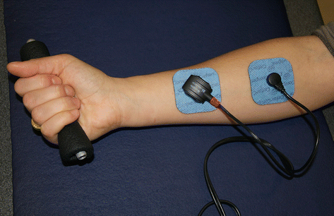
Draining massages without slackening the scar in case of surgery. Decontracting massages for the forearm if we observe hypertonia in the extrinsic musculature, which is frequent (Fig. 3.8).
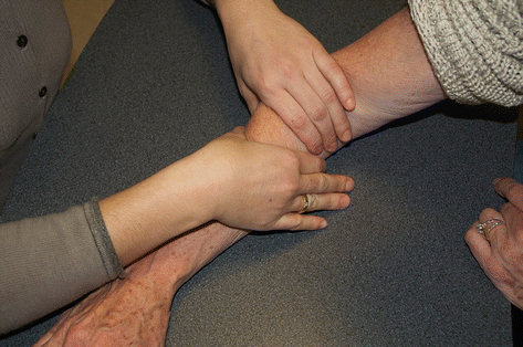
Fig. 3.8
Decontracting massage of the forearm muscles
Analgesic electrotherapy can be used.
Pressotherapy from distal to proximal helps draining the wrist. In this phase, this technique can be badly tolerated by the patient; we’ll then delay its use.
Transcutaneous vibratory stimulations and infrasounds are used for their analgesic and vasomotor effect. Vibrations with more than 1 mm amplitude are realized far from the fracture site in order not to disturb the healing process.
Regaining Functional Amplitudes
It requires a good knowledge of the constraints transmitted to the scaphoid depending on the joint sector [16].
In flexion-extension, the ligaments are relaxed and the joint pressure minimal until 20°, to become significant at 40°. After that, the pressures increase to reach their maximal value around 80°. After 80°, they become pathogenic [6] (Fig. 3.9).
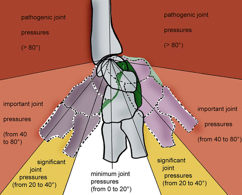
Fig. 3.9
Joint pressures depending on the amplitudes in flexion/extension, which become important after 40° (According to Kapandji)
The normal amplitude in radial inclination is 15°. It induces compressive constraints on the scaphoid that tilts in flexion, which can encourage the formation of a vicious callus [17], closing the intrascaphoidal sagittal angle.
The normal amplitude in ulnar inclination is 45°. It creates traction constraints on the scaphoid that tilts in extension. The formation of a vicious callus [17] when the intrascaphoidal sagittal angle is open is rarer, but can happen.
Like for the fractures of the inferior extremity of the radius, these elements allow defining “risky” areas in which the exercises to regain amplitude will have to be realized very cautiously, without any force.
These areas are after 20° in flexion/extension and ulnar inclination and from 5° in radial inclination.
Trying to regain total amplitudes at all costs is, in this phase, useless and dangerous.
We use the same techniques than in fractures of the inferior extremity of the radius:
Fluidotherapy to relax the muscles and tissues. We use it at the beginning of the session to warm up the wrist.
Passive manual mobilizations are basic exercises to regain joint amplitude. They’re soft and pain-free and can be realized increasing the joint spaces a little. We work in all the authorized sectors, being particularly careful in extension and radial inclination where the constraints on the scaphoid are more important.
Active mobilizations are realized after the passive ones, in various finger positions to improve tendinous glidings at the level of the wrist (Fig. 3.10).
Fig. 3.10
Active mobilizations realized with the fingers extended and a closed fist to improve the tendon glidings in the flexor and extensor systems
Active–assisted mobilizations can be assisted by an arthromotor. We ask the patient to participate actively in the movements applied by the device. This exercise is realized in amplitudes previously regained by the therapist in passive. It has an interest at the joint level, but also in draining the edema. It can be combined with icing (Fig. 3.11 and 3.11′).
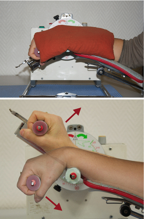
Fig. 3.11 and 3.11′
Active-assisted mobilization with an arthromotor with or without ice depending on the trophic state of the wrist
Electrostimulation, combined with an active movement (winding the fingers), can help regaining the last degrees of mobility in finger flexion if necessary (Fig. 3.12). The patient mustn’t clutch his fingers as it would put important constraints on the scaphoid.