Radiocarpal Dislocation
19.1 Introduction
Radiocarpal dislocations are rare complex injuries whose description and treatment remain confusing. Hippocrates considered that radiocarpal dislocations were the only wrist injury (the joint dislocates either medially, or laterally, most often medially), which explains why the first descriptions of distal radius fracture by Pouteau and Colles were made rather late. A few years later, Dupuytren stated that radiocarpal dislocations were extremely rare or even nonexistent. Even with the use of radiography, their real frequency has still been disputed. Dunn stated that their frequency was 0.2% of all dislocations, while Moneim stated that they could represent 20% of all wrist injuries.1,2 Most of the 70 cases reported in the 19th century, and collected by Abadie, were not radiocarpal dislocations but intracarpal dislocations, epiphyseal injuries, or very displaced wrist fractures.3 Since 1901, few cases of radiocarpal dislocations have been reported, with much confusion. Dunn, who is frequently quoted, reported six cases in 1972.1 However, one of his cases was a very comminuted distal radial fracture, and in the two other reported cases with radiography, one dislocation was secondary to a fracture of the anterior margin (Letenneur fracture), and the other to a fracture of the posterior margin (Barton fracture). Most reports deal with the same problem of confusion. From the review of 438 radiographs, Ilyas et al4 found that radiocarpal dislocation represent 2.7% of patients (12 cases, 10 with associated fracture).
About 80 cases were found in our 1995 review of the literature since Arcelin in 1921.3 PubMed search has found about 30 more publications but they are not all accessible to readers. Since this 1995 review, only three series reported more than eight cases.5–7
These injuries are usually caused by a high-velocity trauma. They are associated with various ligamentous injuries and fracture patterns. Dorsal dislocations are more common than volar dislocations (a 10:2 ratio according to Ilyas, a 23:4 ratio in our series).4,6 The soft tissue disruption can lead to instability, resulting in ulnar translocation and multidirectional radiocarpal instability. As opposed to the reported severity of the injury, most of the results were notably good, especially after orthopedic treatment, whatever the direction of the dislocation. We reported our experience in 2001 comprising 27 cases in which our clinical and radiological results were not as good.6 Since then, others have reported similar less successful results.7,8
19.2 Mechanism of Injury
The exact mechanism of radiocarpal dislocation is still unknown. From a literature review and our own experience of 27 cases, we postulated that the dislocation resulted in a combination of wrist hyperextension, frontal deviation, and rotational movement.6 Abadie, in 1901, was only able to reproduce these lesions with a 180° rotation of the carpus under the radius. Weiss et al9 were able to create a dorsal radiocarpal dissociation with disruption of the distal radioulnar joint by applying compressive and torsional force. The wrist was in a hyperextended and pronated position.
The literature indicates that posterior dislocations are possibly associated with wrist hyperextension, pronation, and radial deviation.10–12 Hyperextension alone is probably unable to produce a radiocarpal dislocation. When an added rotational movement was postulated, the frequency of distal radioulnar joint lesions became an additional argument13 (▶ Fig. 19.1). Dodd is the only author who reported the possibility of a hyperflexion mechanism.3 Frontal inclination was also postulated, but the mechanism of radiocarpal dislocation is probably different from the mechanism of perilunate injuries as intracarpal lesions have scarcely been reported in radiocarpal dislocation.3
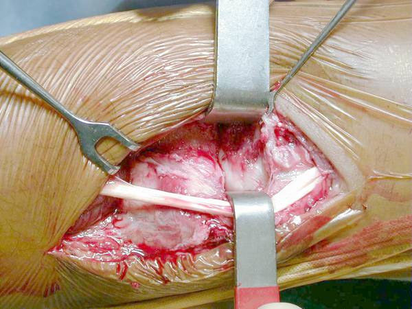
Fig. 19.1 Peroperative view of an avulsion injury of the distal radioulnar joint (DRUJ) in a pure radiocarpal dislocation. Most patients in the literature present with DRUJ lesion suggesting a rotational movement during injury.
In one unpublished case we were able to identify and document the mechanism. A 34-year-old man, a right-handed garage owner, presented with a dorsal radiocarpal dislocation. After a fall from his motorbike, his right hand was trapped between a wall and his motorbike’s wheel. To remove his hand, the patient pulled violently on his arm, resulting in an axial traction. The lesions seen during surgery and arthroscopy were concordant with an axial traction injury. In that case, even the distal radioulnar joint lesion was apparently a traction injury and not a rotational injury.
19.3 Classification
Moneim et al2 proposed a classification for radiocarpal fracture-dislocations into two types, with or without intercarpal ligament injuries (scapholunate or lunotriquetral) in addition to the radiocarpal dislocation.
We have proposed a classification using the type of injury to guide the treatment decision.3,6 Two types of radiocarpal dislocations based on anatomical considerations are described.
Type 1 includes pure ligamentous radiocarpal dislocations or dislocations with only a small cortical or radial styloid avulsion fracture (▶ Fig. 19.2). Anatomical descriptions, experimental studies, literature review, and our own surgical experience show that the radioscaphocapitate and short radiolunate ligaments are torn. This ligamentous tear is sometimes replaced by a chip fracture at the insertion site of the ligaments, or a tiny irreparable radial styloid tip avulsion. Dorsally, the ligamentous injury is most often a capsuloperiosteal avulsion (a “Bankart type” avulsion), rather than a rupture of the dorsal radiocarpal ligament.
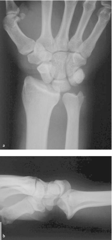
Fig. 19.2 Pure radiocarpal dislocations are rare injuries with avulsion of ligaments from the radius. Most often these are posterior dislocations. (a) AP view; (b) lateral view.
Type 2 includes dislocations with a large radial styloid fracture fragment involving at least one-third of the scaphoid fossa of the distal radius (▶ Fig. 19.3). In our series, the radial styloid fragment was not larger than the scaphoid fossa and the fracture line was always horizontal (which is different from the vertical radial styloid fracture line seen in the greater arc intracarpal injuries).
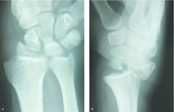
Fig. 19.3 In group 2 radiocarpal dislocations, an associated fracture of the radial styloid is seen. However, in the majority of patients, the fracture line is mostly horizontal and due to rotational injury. (a) AP view; (b) lateral view.
Type 1 injuries are pure ligamentous injuries that have the potential to develop into multidirectional and highly unstable conditions. In type 2, most of the radiocarpal ligaments remain attached to the fractured large radial styloid bony fragment. Stability can more reliably be restored with secure anatomical osteosynthesis of the fracture that can occur with the more pure ligamentous injuries seen in type 1.8
Wang et al13 proposed to add a third type for more complex injury patterns such as the ones where there is an associated injury of the distal radioulnar joint. We prefer to include this specific pattern in addition to the two types described previously.
19.4 Clinical Presentation
Related posts:
 Undisplaced Scaphoid Fractures
 Proximal Row Fractures (Other Than Scaphoid and Pisiform Fractures): Triquetrum and Lunate Fractures
 Intra-articular Fractures of the Distal Radius (AO types C3, with Special Focus in C3.3), Open Approach
 Intra-articular Fractures of the Distal Radius (AO type C3), (Dry) Arthroscopic Approach
 Galeazzi Fracture-dislocation
 Unstable Ulnar Styloid Fracture
Stay updated, free articles. Join our Telegram channel

Full access? Get Clinical Tree


