55 Radial Shortening
Indications
Symptomatic Kienböck disease without lunate collapse (stages 0, I, or II) in a wrist with a positive ulnar variance
Technique
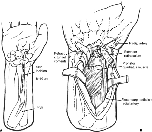
- Palmar incision over the flexor carpi radialis (FCR) tendon beginning at wrist crease and extending 8 to 10 cm up the forearm
- Open FCR tendon sheath and retract tendon in ulnar direction. Incise the floor of the tendon sheath.
- Identify and incise the pronator quadratus (PQ) along its radial margin and elevate from the distal radius (Fig. 55-1A,B).
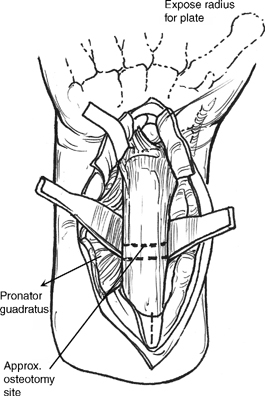
- Place retractors on both sides of the radius. Expose enough of the radius to accommodate a six-hole dynamic compression plate (DCP) (Fig. 55-2). This exposure may require partial elevation of flexor pollicis longus (FPL) origin.
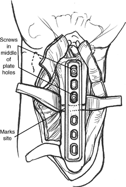
- Apply the six-hole DCP along distal radius. The distal aspect of the plate may have to be bent to match the palmar slope of the distal radius.
- Mark the osteotomy site between third and fourth holes in the plate. Place bicortical screws distal to the planned site of the osteotomy.
Figure 55-1
Figure 55-2
Pearl
The osteotomy should be performed in the metaphysis to increase the chance and rate of healing.
- Remove the screws and plate. Elevate the periosteum at osteotomy site.
- Make the first cut two thirds of the way through the radius. This cut can be either transverse or oblique to the long axis of the radius. A second cut is made parallel to the first, 2 to 4 mm proximal to the initial osteotomy. Complete the first cut and remove wafer of radius.
- Reapply the plate and distal screws. Compress the osteotomy by manual pressure until bone ends approximate. Hold with reduction clamp applied between the proximal plate and radius (Fig. 55-3).
- Secure the proximal portion of the plate using one or two compression screws to compress the osteotomy site. Place the remaining screws in neutral compression (Fig. 55-4).
- Check the alignment of the osteotomized radius and the plate and screw position with fluoroscopy (Fig. 55-5).


