Fig. 22.1
Mason classified radial head fractures as nondisplaced, displaced, and displaced and comminuted [18]. Johnston added a fourth type, which entails all radial head fractures in concomitance with an elbow dislocation
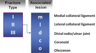
Fig. 22.2
Van Riet and coworkers have proposed a more detailed classification which classifies the associated injuries as well
22.2.1 Isolated Radial Head Fractures
Nondisplaced radial head fractures are amenable to conservative treatment. They will generally prove to be stable enough to allow for early mobilization. They do not give rise to mechanical blocks or residual locking symptoms and will, almost without exception, lead to bony healing within 6 weeks after trauma. Although seemingly trivial injuries, they usually do give rise to a prolonged period of lateral elbow pain, as well as a slight (but sometimes permanent) extension deficit. This might be indicative of an underappreciation of the true severity of any trauma resulting in a radial head fracture, and the patient is better off being given a heads up in advance on the potentially deceptive nature of this seemingly innocuous type of fracture. The long-term results of conservative treatment of undisplaced fractures are, in general, good. It is mostly the displaced partial articular (Mason type 2) fractures that lead to debates whether conservative or operative treatment should be recommended. A commonly used criterium for operative intervention, when considering the appearance of the fracture fragment, is a displacement of more than 2 mm and a size of the fragment entailing more than 30 % of the articular surface. This is also reflected in the classifications as earlier described. There is some evidence that a more conservative approach might be justified [26], and some surgeons only resort to surgical intervention in case of a mechanical block or other absolute indications for surgery. When opting for operative treatment, it can be noted that the fracture fragment is usually located within the anterolateral quadrant of the radial head with the forearm in neutral. This corresponds well to the non-articular part of the rim which allows for placement of screws or other fixation devices as desired without compromising the true articular surface (Fig. 22.3). It should be borne in mind, however, that this also means that, when inadvertently penetrating the overlying cortex, the other end of the screw always ends up damaging the articular surface.
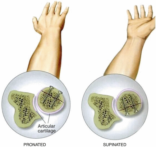
Fig. 22.3
The fracture fragment is usually located within the anterolateral quadrant of the radial head with the forearm in neutral. This corresponds well to the non-articular part of the rim which allows for placement of screws or other fixation devices as desired without compromising the true articular surface
Complete articular fractures with displacement are candidates for surgical intervention and will usually be treated with ORIF or, if not amenable to reconstruction, prosthetic replacement. When the fracture consists of three large fragments, it is usually feasible to reconstruct the articular surface in a satisfactory manner. But even in more comminuted patterns, it might be worthwhile trying to retain the patient’s own joint, when the patient is young and active. If reconstruction is not deemed a viable option, the most common alternative is prosthetic replacement of the radial head.
22.2.2 Associated Injuries
Certain radiographic patterns of fracture may be predictive of the presence of other fractures of the elbow or concomitant ligament injuries, thus representing a complex injury pattern. In a series of 121 Mason type 2 fractures, the presence of a fracture fragment that lacked cortical contact with the rest of the proximal radius was associated with such concomitant lesions in 91 % of cases, while this was 33 % when cortical contact remained [23].
The interobserver agreement of determining the lack or presence of cortical contact using AP and lateral x-rays was reported to be moderate [3]. Future studies may clarify the potential added value of CT scans for this particular purpose.
Another study confirmed in a series of 18 patients who were clinically suspected for longitudinal forearm injury that no lesion of the interosseous membrane was present in Mason 1 radial head fractures, as confirmed by MRI. Both Mason 2 and 3 classified radial head fractures were associated with partial or complete tearing of the interosseous membrane in this small series of clinically suspect patients. It was also noted that a substantial part of patients, even those with an intact interosseous membrane, showed edema in the pronator quadratus muscle. This finding correlated with distal forearm pain.
22.2.3 Elbow Dislocations with Radial Head Fracture
Elbow dislocations with a concommitant radial head fracture are typically part of the terrible triad, in which a coronoid fracture and injury of the lateral ulnar collateral ligament are also present. The negative connotation with this injury, as reflected in its given name, is a remnant of the historically poor results originally obtained when treating these patients. Modern insight in the key elements of this injury and advances in the surgical treatment have led to reproducible and generally good results [22]. It might still be an “unhappy” triad, but calling it “terrible” does not seem to be entirely appropriate these days. Conservative treatment is the exception to the rule in this injury pattern. Prerequisites are a radial head fracture and coronoid fracture type that are both fit for conservative treatment, as well as an elbow which is stable from full flexion to at least 45° of flexion. A cast with the elbow in 90° of flexion and full pronation is applied for the first 10 days, followed by dynamic bracing with a progressively more lenient extension block or a removable splint in 30° of extension. Keeping the forearm in pronation during the first weeks unloads the lateral ulnar collateral ligament, as in pronation the radial head is firmly reduced against the capitellum and creates the best chance of healing of this structure without attenuation.
The mainstay of treating the unhappy triad remains surgical intervention. Preoperative imaging, including a CT scan with 3D reconstruction, is helpful in making a tailor-made plan for the individual patient. When performing the surgery, the injuries are assessed from outside-in, while treatment advances from inside-out. This means that the injury to the lateral ulnar collateral ligament is identified first. It is usually avulsed from the proximal (humeral) attachment. Next, the radial head fracture is assessed. If it is deemed to be reconstructible, reconstruction is first delayed until the coronoid fracture has been addressed.
If the decision is made to replace the radial head by a prosthesis, the resection is done next (but no prosthesis is implanted yet) to enhance visualization of the coronoid fragment. Then, starting from the innermost injury, the stepwise treatment is initiated and the coronoid injury is addressed. If it is a mere flake fragment, it might be left untouched. Preoperative imaging might however underappreciate the true size of the coronoid fragment, and, if feasible, any substantial fragment should be reduced and fixed with either screws, transosseous sutures, or suture anchors. Next, the radial head is either fixed or replaced. Finally the lateral ulnar collateral ligament is reattached to its origin. Postoperative rehabilitation is dictated by the stability of the radial head (prosthesis or osteosynthesis) and the quality of the fixation of the coronoid. In general we apply a plaster for 10 days, replace the plaster by a removable cast, and start mobilization against gravity by a specialized physiotherapist. Axial loading is forbidden for 3–4 months as is forceful lifting in supination.
22.3 Presentation and Clinical Exam
Taking a careful history and taking into account the mechanism of injury may lead to important clues to the severity of injury and potential associated injuries. Although some patients are perfectly able to point out the lateral side of the elbow to be involved in the injury, others are not. A careful examination of the elbow will usually reveal tenderness at the lateral side. Both flexion and extension are usually limited as a consequence of the resulting hemarthrosis. Rotational movements may elicit pain or even a mechanical block.
The latter is regarded a strong indication for surgical treatment.
Even after identifying the radial head as the potential site of injury, the remainder of osseous and ligamentous structures should be assessed as well. A concomitant examination of the entire upper extremity is needed to rule out concommitant injury either proximal or distal to the elbow, such as an Essex-Lopresti forearm dissociation.
22.4 Imaging
Conventional x-rays are the mainstay of confirming the diagnosis of a radial head fracture. In the undisplaced fracture type (Mason 1), the only radiographic clue might be a positive fat pad sign. The anterior fat pad is, especially in younger patients, sometimes also visible in the uninjured elbow, but a visible posterior fat pad is highly suggestive of intra-articular fluid or hemarthrosis.
Displaced or comminuted radial head fractures are usually readily visible, although the exact amount of displacement, the percentage of damaged articular surface, and severity of comminution can be difficult to establish.
When relying on these criteria for determining the definitive treatment, a CT scan may be helpful. Evidence suggests that the addition of 3D CT images can lead to improvement of reliability of classifying radial head fractures according to the Broberg and Morrey modification of the Mason classification. Moreover, it seems to lead to a higher sensitivity to detect radial neck fracture and comminution, articular gaps, step off of at least 2 mm, and impaction of the articular surface. The amount of displacement is often overestimated at standard radiographs, while the comminution is often underestimated [13].
22.5 Treatment Options, Complications, and Outcome
22.5.1 Conservative Treatment
A Cochrane review published in 2014 found two randomized controlled trials comparing aspiration versus no aspiration for treating radial head fractures in adults [10]. The two trials included 126 participants, but results were provided for 108 patients only. One trial included Mason 1, 2, and 3 fractures as well as a few cases with hemarthrosis with clinical suspicion of a fracture, but, besides a positive fat pad sign, no clear radiological sign of fracture after trauma [14]. The other trial included Mason types 1 and 2 fractures only [7]. All cases were treated conservatively. The substantial risk of bias in both studies led the authors of the Cochrane review to downgrade the level of evidence to “very low quality”, concluding that there is insufficient evidence to determine the effectiveness of joint aspiration for the initial treatment of radial head fractures in terms of function, pain, and range of motion or to determine the safety of the procedure.
Related posts:


Stay updated, free articles. Join our Telegram channel

Full access? Get Clinical Tree


