Triquetrum Fractures
8.2.1 Background
Fractures of the os triquetrum are the most common carpal fractures after those of the scaphoid. Triquetral fractures are classified into fractures of the dorsal cortex and fractures of the body: these types differ concerning mechanism of injury, diagnosis, therapy, and prognosis.
8.2.2 Fractures of the Dorsal Cortex of the Triquetrum
Dorsal cortical fractures are rather frequent and make the triquetrum number two of all carpal bones affected by bony injuries. The size of the dorsal fragment may vary from crumbs to a large cortical chip.
Mechanism of Injury
These fractures can result from impaction, avulsion, or shear mechanisms. A fall on the extended hand with the wrist in ulnar deviation is the most frequent causative accident.
Impaction is considered the most common mechanism and may be promoted by a long ulnar styloid. Garcia-Elias measured the size of the ulnar styloid in 76 patients with dorsal fractures of the triquetrum and compared the results with the measurements in 100 noninjured hands. The size of the ulnar styloid was found to be significantly larger in the fracture group (P < 0.0001).5 This finding supports the concept that during strong dorsiflexion the ulnar styloid process may act as a chisel and lead to dorsal fractures of varying extent.
Shear forces may be applied during hyperextension by the hamate to the distal dorsal part of the triquetrum.6 This mechanism was favored by Höcker and Menschik,7 who analyzed a series of 63 chip fractures.
In cases of avulsion, dorsal fractures would represent a bony detachment of the dorsal radiotriquetral or intercarpal ligament.
Diagnosis
Physical examination usually shows uncharacteristic symptoms such as moderate swelling and a painful limitation of the range of wrist motion. Sometimes skin abrasion and hematoma on the palmar site suggest a hyperextension injury. Palpation reveals maximal tenderness over the dorsal aspect of the triquetrum and the ulnocarpal joint space.
Plain radiographs of the wrist in two routine views (postero-anterior [PA], lateral) should primarily exclude other injuries to the carpus or distal radius such as displaced fractures or dislocations. Whereas fractures of the dorsal cortex of the triquetrum can rarely be detected on PA views, they are often already visible on lateral views (▶ Fig. 8.1a, b). More helpful are additional oblique views with the wrist in semipronation. In all cases of uncertainty or suspected other carpal injuries, computed tomography is strongly recommended. Primary axial scans should be obtained at 0.5- to 0.75-mm intervals. Postprocessing should include multiplanar reconstructions in coronal and sagittal plane. Three-dimensional reconstructions are optional. Computed tomography is especially helpful in assessing the size and location of the fragment as well as its displacement (▶ Fig. 8.1c, d).
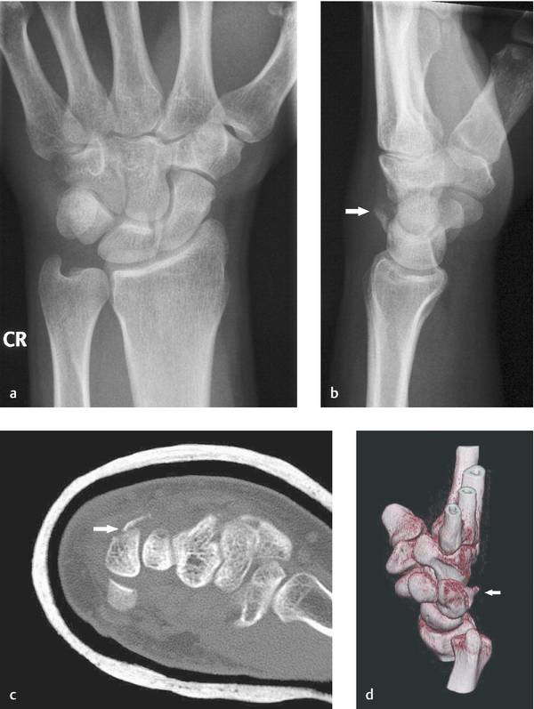
Fig. 8.1 Dorsal cortical fracture of the triquetrum (chip fracture). (a and b) On plain radiographs, the fragment is visible on the lateral view. (c and d) CT scans demonstrate size, location, and displacement of the fragment.
MRI can provide additional information on the involvement of ligaments but is not expected to change treatment decisions. In their MRI study on 21 patients with dorsal fractures of the triquetrum, Becce et al8 found tears of the dorsal radiocarpal, ulnotriquetral, and intercarpal ligaments in 67%, 81%, and 76%, respectively.
Classification
Based on their analysis of 231 triquetrum fractures, Höcker and Menschik7 proposed the following classification of chip fractures that is intended to be only of descriptive value:
Not displaced
Completely displaced
Partial displacement—proximal end
Partial displacement—distal end
Multiple fragments
Treatment
Routinely, cortical fractures of the dorsal aspect of the triquetrum can be treated conservatively by splint or cast immobilization of the wrist until the pain has subsided. Two to three weeks of immobilization are sufficient in most cases. If pain persists for more than 4 weeks, other relevant causes should be excluded. Cortical fractures that represent avulsions of the dorsal ligaments may also be treated conservatively but require prolonged splinting for 6 weeks.9
Prognosis
The dorsal fragment often remains ununited but this rarely causes symptoms. Displacement of a fragment of 2 mm or more promotes nonunion and formation of an ossicle.7 Only if there is persistent pain over months, excision of the tender fragment should be offered to the patient.6,10 In their series of 63 chip fractures, Höcker and Menschik7 saw no case that needed surgical intervention and did not observe signs of carpal instability as well.
Lee et al11 performed MRI and wrist arthroscopy in six patients with ongoing ulnocarpal joint pain and tenderness after triquetral dorsal chip fracture and in all six cases found triangular fibrocartilage complex (TFCC) injury, which was treated by partial TFCC resection.
Summary
Dorsal cortical fractures of the triquetrum are the most common carpal fractures after those of the scaphoid. They result from ulnar styloid impaction or shear mechanisms and can be diagnosed in most cases by plain radiography. Treatment is preferentially nonsurgical with wrist immobilization for up to 3 weeks. Although these chip fractures often remain ununited, prognosis is generally good and surgical measures are seldom required.
8.2.3 Fractures of the Body of the Triquetrum
Fractures of the body represent the other main bony injury to the triquetrum but are less frequent. Of 231 triquetrum fractures that were analyzed by Höcker and Menschik7 only 3% were fractures of the body. These fractures usually occur as a part of perilunate fracture-dislocations. Isolated triquetrum body fractures are typically undisplaced.
Mechanism of Injury
Whereas the trauma pattern for fractures of the dorsal corticalis has been investigated and discussed in several studies, the mechanisms leading to body fractures are not well explained. Aside from perilunate fracture-dislocations with distribution of forces from radial to ulnar and involving the triquetrum, body fractures may be best explained by forces resulting from direct trauma at the ulnar side of the wrist. In most circumstances, this is the result from a fall on the extended wrist. The pisiform can transform forces to the triquetrum, or may be affected by fracture or ligamentous injury itself. Another mechanism could be direct trauma by the ulnar styloid in hyperextension and ulnar deviation, which can again act as a chisel but—depending on the position of the bones and the vector of the transmitted forces—lead not to a cortical but to a body fracture.
Diagnosis
In the absence of fracture-dislocation, symptoms may be minor but physical examination will show swelling and painful limitation of wrist motion. Palmar skin laceration and hematoma can indicate a hyperextension injury. Tenderness during palpation and motion can be found both on the dorsoulnar aspect of the wrist and palmar over the pisiform.
Related posts:


Stay updated, free articles. Join our Telegram channel

Full access? Get Clinical Tree


