45 Proximal Row Carpectomy with Capsular Resurfacing
Indications
- Radioscaphoid arthritis with preservation of the radiolunate and midcarpal joints. This pattern is typically seen in long-standing scapholunate dissociation.
Pitfall
Degenerative changes on the capitate may not be apparent on the preoperative radiographs. Patients should be warned about the possibility of an alternative procedure, such as a scaphoid (S) excision and capitate—lunate—triquetral—hamate (CLTH) fusion.
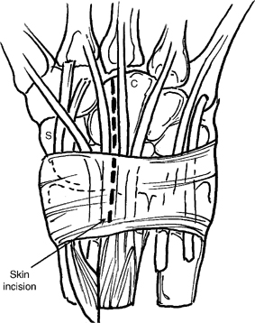
Figure 45-1
Technique
- A dorsal, longitudinal, 6 to 8 cm skin incision is centered over the capitate (C) (Fig. 45-1).
- Release the extensor pollicis longus from the third compartment and retract radially.
- Incise the radial septum of the fourth compartment, elevating the retinaculum as a flap.
- Retract the radial wrist extensors in a radial direction and the extensor digitorum communis in an ulnar direction.
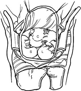
- Create a U-shaped, distally based capsular flap (Fig. 45-2).
Figure 45-2
If unexpected degenerative changes are found on the head of the capitate, the capsular flap can be interposed between the capitate and the lunate (L) fossa.
Pitfall
The flap must be wide enough to completely cover the head of the capitate. The radial margin should be raised beneath the extensor carpi radialis brevis tendon (ECRB); the ulnar margin should be raised just radial to the fifth extensor compartment.
- Inspect the articular surfaces of the capitate and lunate fossa.
- Wear on the capitate is handled in two fashions:
- Young patient, nonsmoker: scaphoid excision and CLTH fusion.
- Older patient, smoker: proximal row carpectomy with interposition of the capsular flap.
- Split the scaphoid across its waist with an osteotome and remove the proximal pole of the scaphoid.
- Split remaining distal pole of the scaphoid along its longitudinal axis and remove both pieces with a rongeur.
Pitfall
Avoid injury to the radioscaphocapitate ligament passing palmar to the scaphoid waist.
- Excise osteophytes from the radial styloid.
- Drill a heavy, threaded pin into the lunate and use as a joystick to help remove the lunate with sharp dissection.
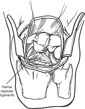
- Remove the triquetrum using the same technique as described for removal of the lunate (Fig. 45-3).
- If you chose to interpose the capsule, use a 2–0 suture on a short arc needle to suture the dorsal flap to the palmar capsule.
- Use three horizontal mattress sutures with the knot tied dorsally by passing the needle from the dorsal flap through the palmar capsule and back up through the dorsal flap (Fig. 45-4A,B).
- If the cartilage on the head of the capitate is intact close the capsule, retinaculum, and skin.


