and Ulna Fractures
Andrew R. Evans
Daphne M. Beingessner
Sterile Instruments/Equipment
Proximal Ulna Fractures
- Dental picks
- Freer elevators
- Small Hohmann retractors (narrow and wide)
- Small point-to-point clamps
- Kirschner wires
- Implants:
- 3.5/2.7-mm head screws
- 2.0- and 2.4-mm plate and screw sets
- Proximal ulnar periarticular plating sets
- 18-gauge stainless steel cerclage wire or cable
- 5.0-, 5.5-, 6.0-, or 6.5-mm cancellous intramedullary screw
- 3.5/2.7-mm head screws
Radial Head and Neck Fractures
- Dental picks
- Small point-to-point clamps
- Kirschner wires
- Implants:
- 2.0- and 2.4-mm screw sets
- Mini-fragment plate/screw set with T- and L-shaped plates
- Radial head arthroplasty system of choice
- 2.0- and 2.4-mm screw sets
Terrible Triad Injuries of the Elbow
- Dental picks
- Small point-to-point clamps
- Kirschner wires
- In-line ACL drill guide
- Implants:
- 2.0- and 2.4-mm screw sets
- Mini-fragment plate/screw set with T- and L-shaped plates
- Nonabsorbable suture material (Ticron or Fiberwire)
- Radial head arthroplasty system of choice
- Suture anchors
- 2.0- and 2.4-mm screw sets
Surgical Approaches
- Dorsal extensile approach to the proximal ulna.
- For Monteggia fracture/dislocations, comminuted proximal ulna fractures, and olecranon fractures
- Lateral or prone position
- Drape affected arm over radiolucent arm board with the shoulder in a forward flexed (~90 degrees) and partially abducted (~90 degrees) position.
- Verify that adequate C-arm imaging is possible prior to draping (Fig. 7-1).
- The prone position provides improved C-arm access and imaging over the lateral position as there are fewer anatomical and positioning impediments (e.g., the opposite extremity, metal arm board support).
- Drape affected arm over radiolucent arm board with the shoulder in a forward flexed (~90 degrees) and partially abducted (~90 degrees) position.
- For Monteggia fracture/dislocations, comminuted proximal ulna fractures, and olecranon fractures
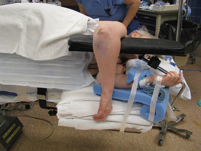
Figure 7-1. Lateral positioning for a proximal ulna fracture.
![]()
- Skin incision located directly over subcutaneous border of the ulna, deviating radial to the olecranon process, and returning to the dorsal midline.
- Avoids dissection of the olecranon bursa.
- Places incision away from contact with desks and tables when extremity is resting on these surfaces.
- Avoids dissection of the olecranon bursa.
- Create full-thickness skin flaps radially and ulnarly to visualize the proximal ulnar shaft and olecranon process.
- If visualization of the medial aspect of greater sigmoid notch (semilunar fossa) is required, the ulnar nerve should be identified in the cubital tunnel and protected.
- Ensure that the path of the ulnar nerve is known, either by palpation or direct visualization.1
- Avoid dissection of soft-tissue attachments to fracture fragments.
- Posterolateral (Kocher) approach to the elbow joint.
- For terrible triad injuries and radial head fractures, and posterolateral elbow dislocations.
- Supine position
- Radiolucent hand or arm table.
- Shift patient to the edge of the table so that elbow rests away from the junction of the hand table and OR table.
- Prep and drape the entire involved upper extremity.
- Be sure that the patient has adequate shoulder abduction and external rotation if access to the medial joint is required.
- Radiolucent hand or arm table.
- Two possible skin incisions to access the deep Kocher interval
- Obliquely across the radial head and radiocapitellar joint.
- Posterior midline incision used with distal skin incision located directly over the subcutaneous border of the ulna.
- Deviate radially around the tip of the olecranon.
- Return incision to central dorsal location just proximal to lateral epicondyle.
- Elevate a full thickness flap laterally (radially), sufficiently to locate the fascial septum separating the extensor carpi ulnaris and anconeus, and incise the fascia sharply just ulnar to the septum.
- Deviate radially around the tip of the olecranon.
- Obliquely across the radial head and radiocapitellar joint.
- Gently elevate anconeus off of the fascial septum, which often leads directly into the elbow joint if the capsule has been traumatically compromised due to the dislocation.
- In terrible triad injuries, the lateral humeral epicondyle will often be “bald” due to avulsion of the lateral ligament complex.
- The superficial fascial layer may be intact and thus the injury may appear to have an intact lateral ligament complex.
- Raising full thickness flaps will ensure that the full extent of the deep soft tissue injury is appreciated.
- The superficial fascial layer may be intact and thus the injury may appear to have an intact lateral ligament complex.
- Identify the annular ligament, lateral ulnar collateral ligament, and the origin of the mobile extensor mass.
- For terrible triad injuries and radial head fractures, and posterolateral elbow dislocations.
Reduction and Implant Techniques
Radial Head Fractures
- Locate all fractured fragments of the radial head (reassemble to confirm restoration of full head circumferentially).
- Check olecranon, radial, and capitellar fossae for any unaccounted fragments, or other fracture debris.
- Take care while retracting distally over the radial neck and keep the forearm pronated.
- This relaxes the posterior interosseous nerve (PIN) and rotates it into a safer position.
- Do not place Hohmann retractors blindly around the radial neck or retract excessively, especially anteromedially.
- This relaxes the posterior interosseous nerve (PIN) and rotates it into a safer position.
- If radial head is multifragmentary and fragments are displaced, remove displaced fragments and place on the back table in a saline moistened gauze or saline solution.
- On the back table, attempt to reconstruct the radial head fragments such that they may either be stabilized to the intact portion of the radial head/neck, or sized appropriately for radial head arthroplasty.
- Reassembling the radial head also confirms that all fracture fragments have been identified (Fig. 7-2).
- Check olecranon, radial, and capitellar fossae for any unaccounted fragments, or other fracture debris.
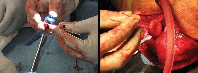
Figure 7-2. The radial head is reassembled either in preparation for arthroplasty replacement (left), or for reduction and fixation (right).
![]()
- Determination of the height of the radial head implant should be made based upon AP and lateral fluoroscopic views of the elbow confirming that the articular surfaces of the coronoid (at the level of the trochlear groove) and radial head are at the same level.
- On the AP view, the medial half of the ulnotrochlear joint should be concentric to avoid “overstuffing.”
- The joint space across the ulnohumeral articulation should be symmetric, accounting for the thickness of the articular cartilage on the coronoid portion of the greater sigmoid notch (Fig. 7-3).
- On the AP view, the medial half of the ulnotrochlear joint should be concentric to avoid “overstuffing.”
- When the patient is supine, positioning of the arm for posterolateral access to the radial head causes an obligatory varus moment across the elbow.
- This can improve access, but care must be taken to neutralize this stress when assessing the radial head height relative to the capitellum.
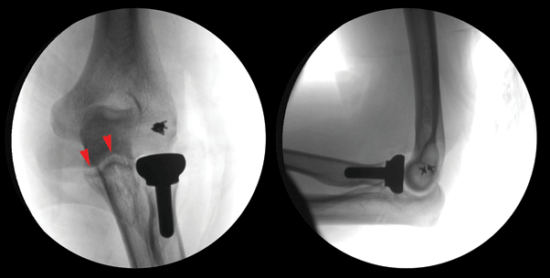
Figure 7-3. Radial head arthroplasty. To ensure that the radiocapitellar joint is not “overstuffed,” the medial ulnotrochlear joint should be symmetric (left, arrowheads).
![]()
- The indication for radial head fixation versus arthroplasty is based upon intraoperative assessment of comminution, the ability to achieve an anatomic reduction with stable fixation of fracture fragments, and overall stability of radial head within the elbow joint.
- In the setting of gross elbow instability associated with radial head fracture, secure fixation of the radial head is mandatory to maintain elbow stability during rehabilitation.
- If it cannot be achieved, then radial head arthroplasty should be performed.
- In the setting of gross elbow instability associated with radial head fracture, secure fixation of the radial head is mandatory to maintain elbow stability during rehabilitation.
Related posts:


Stay updated, free articles. Join our Telegram channel

Full access? Get Clinical Tree


