Proximal Hamstring Injury
Thomas J. Kremen Jr.
Robert T. Sullivan
William E. Garrett
ANATOMY
The hamstring muscle group consists of three muscles: the biceps femoris (long and short heads), the semitendinosus, and the semimembranosus. All three muscles, except for the short head of the biceps femoris, originate from the ischial tuberosity of the pelvis.
The biceps femoris and semitendinosus have a common origin.
The hamstrings are biarticular muscles bridging the hip and the knee.
The proximal tendons of the biceps femoris and semimembranosus have been shown to extend for about 62% and 73%, respectively, of their muscle bellies.
The sciatic nerve lies immediately lateral to the hamstring origin.
PATHOGENESIS
NATURAL HISTORY
The natural history of hamstring strains can be quite different. With a more proximal injury, there is longer time for recovery to preinjury status and a greater likelihood of surgical intervention due to the persistent and significant disability associated with hamstring avulsion.
Partial or complete hamstring avulsions should not be confused with strain at the MTJ. Avulsions can be extremely disabling and, unlike strain at the MTJ, may warrant surgical intervention. Avulsions cause symptoms of weakness and loss of muscle control, especially during running.
Strains most often occur in the biceps femoris and are most commonly located at the proximal MTJ. Fortunately, most proximal hamstring MTJ injuries are best managed nonoperatively. Recovery time has been correlated directly with the percentage of muscle involved by measuring the crosssectional area or the longitudinal length of abnormal muscle signal on magnetic resonance imaging (MRI).2,7,15,23
Injuries involving over 50% of the cross-sectional area result in a recovery period longer than 6 weeks, whereas normal imaging findings result in a recovery period of approximately 1 week.15
The greatest risk factor for injury to the hamstring muscle complex is a history or previous injury to the same place.26,18
Petersen and Hölmich19 reported the recurrence rate for hamstring injury to be 12% to 31%. Whether the reinjury is attributed to insufficient rehabilitation and early return to sport or the persistence of preexisting risk factors, the treating physician must have the ability to assess the degree of injury, knowledge of the reparative process of healing muscle, and an understanding of the rehabilitative and preventive measures for hamstring injury.
PATIENT HISTORY AND PHYSICAL FINDINGS
Proximal hamstring injury typically results in sudden onset of pain in the posterior proximal thigh during athletic competition or training.
Mechanical symptoms, snapping and popping, are not usually reported.
Sports commonly associated with hamstring injury include hurdling, long jumping, sprinting, and waterskiing.
Severe injury, such as an avulsion, may present with a visible deformity, swelling, ecchymosis, and a palpable defect. Focal tenderness to palpation and pain on provocation with resisted knee flexion are consistent findings.
With the patient lying prone and the hamstrings activated, palpation of proximal hamstring origin is undertaken.
A palpable defect implies proximal avulsion. There may be marked asymmetry of the posterior thigh shape.
Pain without a defect suggests partial avulsion.
An apparent muscle-tendon junction sprain can be unmasked by having the patient sequentially contract and relax the hamstrings complex against resistance repeatedly. This assessment can be useful after acute or chronic injury as well as during the return-to-sport phase of rehabilitation.
Obvious increase in apparent hamstring flexibility of the injured extremity implies proximal avulsion.
The current classification of muscle injuries identifies mild, moderate, and severe injuries based on the degree of clinical impairment.
Mild muscle injury is minimal to no loss of strength.
Moderate injury is a clear loss of strength.
IMAGING AND OTHER DIAGNOSTIC STUDIES
Plain radiographs are useful in evaluating for a bony avulsion.
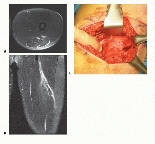
MRI is the imaging of choice to confirm the existence of a muscle injury or avulsion, particularly when a discrepancy exists between the examiner’s findings and the patient’s symptoms.
Despite recent growing interest in the use of ultrasound imaging, it has been demonstrated that proximal hamstring injuries continue to be best imaged with MRI.27
In separate investigations, Connell, Koulouris, Askling, and Slavotinek correlated rehabilitation time to the percentage of muscle involved by measuring the cross-sectional area or the longitudinal length of abnormal muscle signal on MRI.
Schneider-Kolsky et al22 questioned the ability of MRI to predict rehabilitation time for minor and moderate injury. Despite a limitation of variable methods of rehabilitation, they concluded MRI to be useful in predicting the duration of convalescence for moderate and severe injury. Conversely, they determined clinical assessment to be slightly better than MRI for minor injury.
Verall et al26 reported on a subset of patients with a clear clinical diagnosis of hamstring strain but an MRI-negative for muscle injury. Like Schneider-Kolsky et al’s findings, Verall et al26 reported patients with hamstring muscle strain injuries demonstrable by MRI had a poorer prognosis than those whose posterior thigh injury was not MRI-positive.
Askling et al2 also demonstrated a correlation between the location of injury by MRI and time to recovery. They found longer recovery times for injuries in close proximity to the hamstring origin. In other words, the more proximal the injury, the longer the recovery time. Interestingly, the prediction of recovery time was equally good using the point of highest pain on palpation, established within 3 weeks of the injury.
Although not specific to proximal injuries, recently, traditional MRI grading of muscle strain based solely on crosssectional area was compared with a new MRI scoring system taking into account many of the features previously implicated by other authors. These included patient age, location of injury, longitudinal length of abnormal signal, retraction, the number of muscles involved as well as other features. Both the traditional and newly described scoring system correlated with time to return to play among professional American football players.6
Stay updated, free articles. Join our Telegram channel

Full access? Get Clinical Tree


